


 Foundational characteristics of cancer include proliferation, angiogenesis, migration, evasion of apoptosis, and cellular immortality. Find key markers for these cellular processes and antibodies to detect them.
 The SUMOplot™ Analysis Program predicts and scores sumoylation sites in your protein. SUMOylation is a post-translational modification involved in various cellular processes, such as nuclear-cytosolic transport, transcriptional regulation, apoptosis, protein stability, response to stress, and progression through the cell cycle.
 The Autophagy Receptor Motif Plotter predicts and scores autophagy receptor binding sites in your protein. Identifying proteins connected to this pathway is critical to understanding the role of autophagy in physiological as well as pathological processes such as development, differentiation, neurodegenerative diseases, stress, infection, and cancer.
Anti-FACL4/ACSL4 Antibody Picoband™ (monoclonal, 4I7)
- SPECIFICATION
- CITATIONS
- PROTOCOLS
- BACKGROUND

Application
| WB, IHC, IF, ICC, FC |
|---|---|
| Primary Accession | O60488 |
| Host | Mouse |
| Isotype | Mouse IgG1 |
| Reactivity | Human |
| Clonality | Monoclonal |
| Format | Lyophilized |
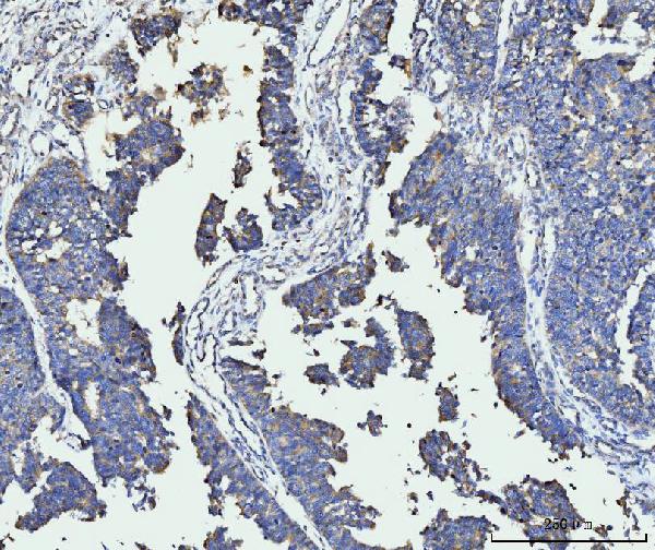
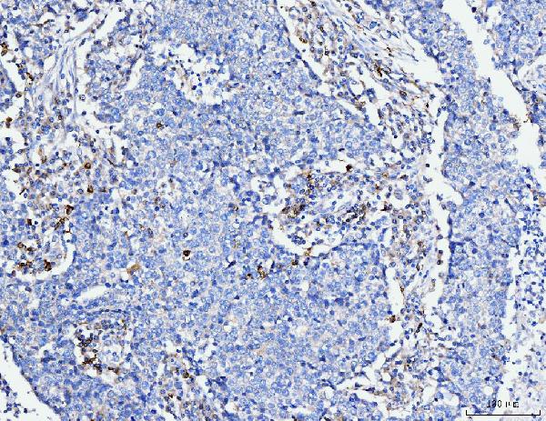
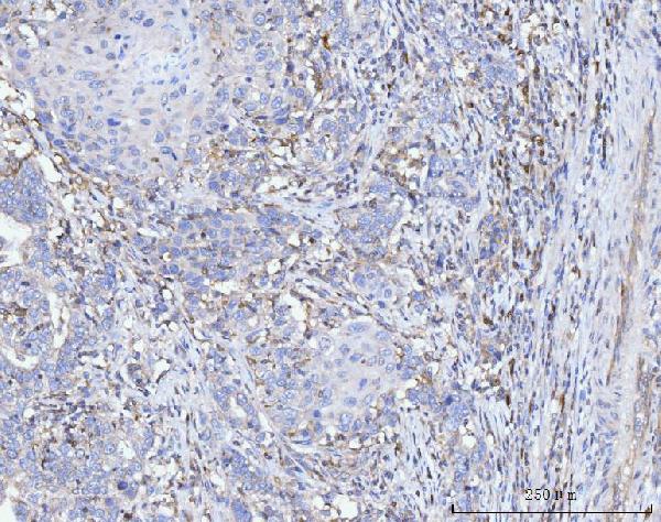
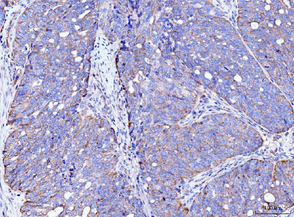
| Description | Anti-FACL4/ACSL4 Antibody Picoband™ (monoclonal, 4I7) . Tested in Flow Cytometry, IF, IHC, ICC, WB applications. This antibody reacts with Human. |
| Reconstitution | Adding 0.2 ml of distilled water will yield a concentration of 500 µg/ml. |
| Gene ID | 2182 |
|---|---|
| Other Names | Long-chain-fatty-acid--CoA ligase 4, 6.2.1.3, Arachidonate--CoA ligase, 6.2.1.15, Long-chain acyl-CoA synthetase 4, LACS 4, ACSL4, ACS4, FACL4, LACS4 |
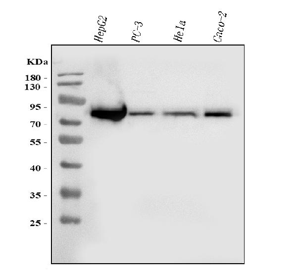
| Calculated MW | 79 kDa |
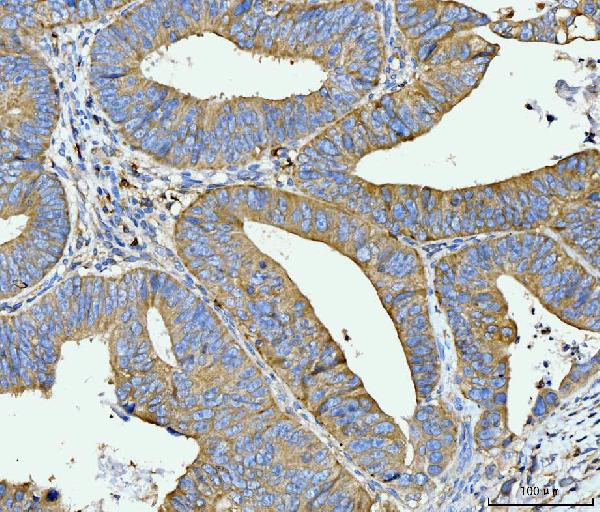
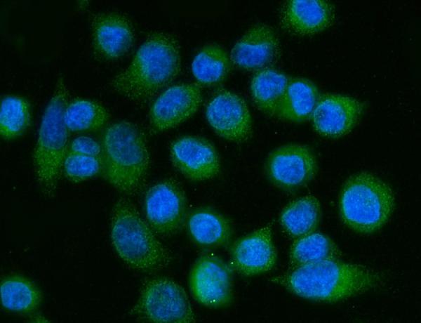
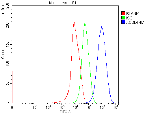
| Application Details | Western blot, 0.25-0.5 µg/ml, Human Immunohistochemistry(Paraffin-embedded Section), 2-5 µg/ml, Human Immunocytochemistry/Immunofluorescence, 5 µg/ml, Human Flow Cytometry, 1-3 µg/1x10^6 cells, Human |
| Contents | Each vial contains 4 mg Trehalose, 0.9 mg NaCl and 0.2 mg Na2HPO4. |
| Clone Names | Clone: 4I7 |
| Immunogen | A synthetic peptide corresponding to a sequence at the C-terminus of human FACL4/ACSL4. |
| Purification | Immunogen affinity purified. |
| Storage | At -20°C for one year from date of receipt. After reconstitution, at 4°C for one month. It can also be aliquotted and stored frozen at -20°C for six months. Avoid repeated freezing and thawing. |
| Name | ACSL4 |
|---|---|
| Synonyms | ACS4, FACL4, LACS4 |
| Function | Catalyzes the conversion of long-chain fatty acids to their active form acyl-CoA for both synthesis of cellular lipids, and degradation via beta-oxidation (PubMed:21242590, PubMed:22633490, PubMed:24269233). Preferentially activates arachidonate and eicosapentaenoate as substrates (PubMed:21242590). Preferentially activates 8,9-EET > 14,15-EET > 5,6-EET > 11,12-EET. Modulates glucose- stimulated insulin secretion by regulating the levels of unesterified EETs (By similarity). Modulates prostaglandin E2 secretion (PubMed:21242590). |
| Cellular Location | Mitochondrion outer membrane; Single-pass type III membrane protein. Peroxisome membrane; Single-pass type III membrane protein. Microsome membrane; Single-pass type III membrane protein. Endoplasmic reticulum membrane; Single-pass type III membrane protein. Cell membrane |

Thousands of laboratories across the world have published research that depended on the performance of antibodies from Abcepta to advance their research. Check out links to articles that cite our products in major peer-reviewed journals, organized by research category.
info@abcepta.com, and receive a free "I Love Antibodies" mug.

Provided below are standard protocols that you may find useful for product applications.
Background
Long-chain-fatty-acid—CoA ligase 4 is an enzyme that in humans is encoded by the ACSL4 gene. It is mapped to Xq23. The protein encoded by this gene is an isozyme of the long-chain fatty-acid-coenzyme A ligase family. Although differing in substrate specificity, subcellular localization, and tissue distribution, all isozymes of this family convert free long-chain fatty acids into fatty acyl-CoA esters, and thereby play a key role in lipid biosynthesis and fatty acid degradation. This isozyme preferentially utilizes arachidonate as substrate. The absence of this enzyme may contribute to the cognitive disability or Alport syndrome. Alternative splicing of this gene generates multiple transcript variants.
If you have used an Abcepta product and would like to share how it has performed, please click on the "Submit Review" button and provide the requested information. Our staff will examine and post your review and contact you if needed.
If you have any additional inquiries please email technical services at tech@abcepta.com.
Ordering Information
Other Products
Shipping Information
