


 Foundational characteristics of cancer include proliferation, angiogenesis, migration, evasion of apoptosis, and cellular immortality. Find key markers for these cellular processes and antibodies to detect them.
 The SUMOplot™ Analysis Program predicts and scores sumoylation sites in your protein. SUMOylation is a post-translational modification involved in various cellular processes, such as nuclear-cytosolic transport, transcriptional regulation, apoptosis, protein stability, response to stress, and progression through the cell cycle.
 The Autophagy Receptor Motif Plotter predicts and scores autophagy receptor binding sites in your protein. Identifying proteins connected to this pathway is critical to understanding the role of autophagy in physiological as well as pathological processes such as development, differentiation, neurodegenerative diseases, stress, infection, and cancer.
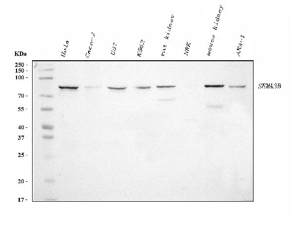
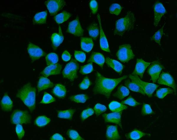
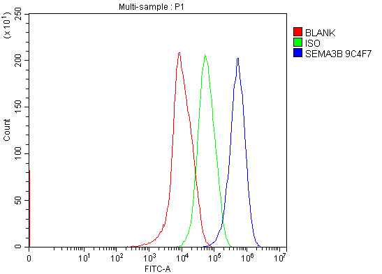
Anti-Semaphorin 3B/SEMA3B Antibody Picoband™ (monoclonal, 9C4F7)
- SPECIFICATION
- CITATIONS
- PROTOCOLS
- BACKGROUND

Application
| WB, IF, ICC, FC |
|---|---|
| Primary Accession | Q13214 |
| Host | Mouse |
| Isotype | Mouse IgG2b |
| Reactivity | Rat, Human, Mouse |
| Clonality | Monoclonal |
| Format | Lyophilized |
| Description | Anti-Semaphorin 3B/SEMA3B Antibody Picoband™ (monoclonal, 9C4F7) . Tested in Flow Cytometry, IF, ICC, WB applications. This antibody reacts with Human, Mouse, Rat. |
| Reconstitution | Adding 0.2 ml of distilled water will yield a concentration of 500 µg/ml. |
| Gene ID | 7869 |
|---|---|
| Other Names | Semaphorin-3B, Sema A(V), Semaphorin-V, Sema V, SEMA3B, SEMA5, SEMAA |
| Calculated MW | 83 kDa |
| Application Details | Western blot, 0.25-0.5 µg/ml, Human, Mouse, Rat Immunocytochemistry/Immunofluorescence, 5 µg/ml, Human Flow Cytometry, 1-3 µg/1x10^6 cells, Human |
| Contents | Each vial contains 4 mg Trehalose, 0.9 mg NaCl and 0.2 mg Na2HPO4. |
| Clone Names | Clone: 9C4F7 |
| Immunogen | E.coli-derived human Semaphorin 3B/SEMA3B recombinant protein (Position: Y55-R624). |
| Purification | Immunogen affinity purified. |
| Storage | At -20°C for one year from date of receipt. After reconstitution, at 4°C for one month. It can also be aliquotted and stored frozen at -20°C for six months. Avoid repeated freezing and thawing. |
| Name | SEMA3B |
|---|---|
| Synonyms | SEMA5, SEMAA |
| Function | Inhibits axonal extension by providing local signals to specify territories inaccessible for growing axons. |
| Cellular Location | Secreted. Endoplasmic reticulum. Note=Accumulates in the endoplasmic reticulum |
| Tissue Location | Expressed abundantly but differentially in a variety of neural and nonneural tissues |

Thousands of laboratories across the world have published research that depended on the performance of antibodies from Abcepta to advance their research. Check out links to articles that cite our products in major peer-reviewed journals, organized by research category.
info@abcepta.com, and receive a free "I Love Antibodies" mug.

Provided below are standard protocols that you may find useful for product applications.
Background
Semaphorin-3Bis aproteinthat in humans is encoded by theSEMA3Bgene. It is mapped to 3p21.31. The semaphorin/collapsin family of molecules plays a critical role in the guidance of growth cones during neuronal development. The secreted protein encoded by this gene family member is important in axonal guidance and has been shown to act as a tumor suppressor by inducing apoptosis.
If you have used an Abcepta product and would like to share how it has performed, please click on the "Submit Review" button and provide the requested information. Our staff will examine and post your review and contact you if needed.
If you have any additional inquiries please email technical services at tech@abcepta.com.
Ordering Information
Other Products
Shipping Information
