


 Foundational characteristics of cancer include proliferation, angiogenesis, migration, evasion of apoptosis, and cellular immortality. Find key markers for these cellular processes and antibodies to detect them.
 The SUMOplot™ Analysis Program predicts and scores sumoylation sites in your protein. SUMOylation is a post-translational modification involved in various cellular processes, such as nuclear-cytosolic transport, transcriptional regulation, apoptosis, protein stability, response to stress, and progression through the cell cycle.
 The Autophagy Receptor Motif Plotter predicts and scores autophagy receptor binding sites in your protein. Identifying proteins connected to this pathway is critical to understanding the role of autophagy in physiological as well as pathological processes such as development, differentiation, neurodegenerative diseases, stress, infection, and cancer.
Anti-Glutaminase/GLS Antibody Picoband™ (monoclonal, 3G13)

- SPECIFICATION
- CITATIONS
- PROTOCOLS
- BACKGROUND

Application
| WB, IHC, IF, ICC, FC |
|---|---|
| Primary Accession | O94925 |
| Host | Mouse |
| Isotype | Mouse IgG2a |
| Reactivity | Rat, Human, Mouse |
| Clonality | Monoclonal |
| Format | Lyophilized |
| Description | Anti-Glutaminase/GLS Antibody Picoband™ (monoclonal, 3G13) . Tested in Flow Cytometry, IF, IHC, ICC, WB applications. This antibody reacts with Human, Mouse, Rat. |
| Reconstitution | Adding 0.2 ml of distilled water will yield a concentration of 500 µg/ml. |
| Gene ID | 2744 |
|---|---|
| Other Names | Glutaminase kidney isoform, mitochondrial, GLS, 3.5.1.2, K-glutaminase, L-glutamine amidohydrolase, Glutaminase kidney isoform, mitochondrial 68 kDa chain, GLS, GLS1, KIAA0838 |
| Calculated MW | 56-73 kDa |
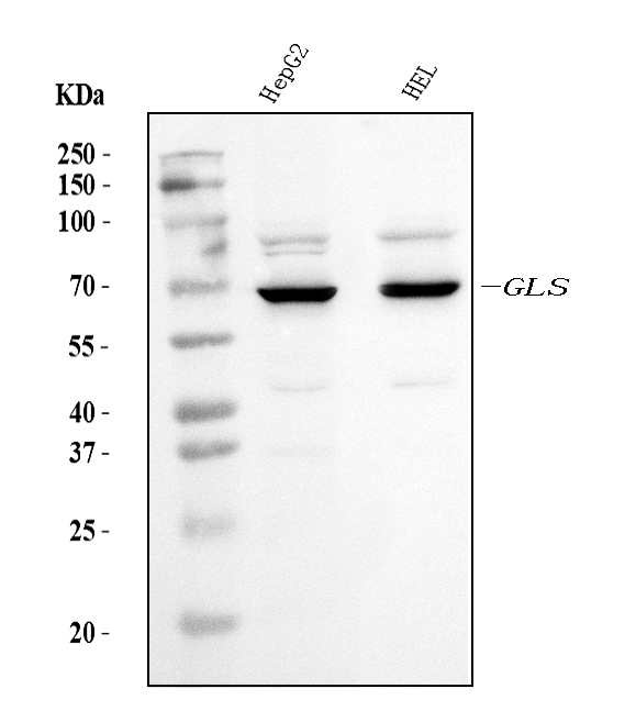
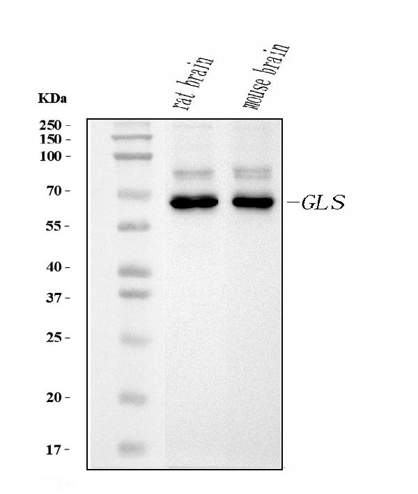
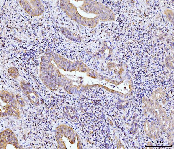
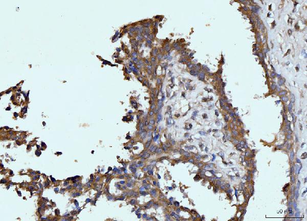
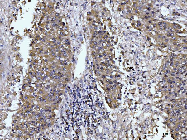
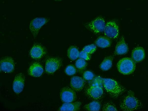
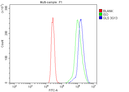
| Application Details | Western blot, 0.25-0.5 µg/ml, Human, Mouse, Rat Immunohistochemistry(Paraffin-embedded Section), 2-5 µg/ml, Human Immunocytochemistry/Immunofluorescence, 5 µg/ml, Human Flow Cytometry, 1-3 µg/1x10^6 cells, Human |
| Contents | Each vial contains 4 mg Trehalose, 0.9 mg NaCl and 0.2 mg Na2HPO4. |
| Clone Names | Clone: 3G13 |
| Immunogen | E.coli-derived human Glutaminase/GLS recombinant protein (Position: K396-N654). |
| Purification | Immunogen affinity purified. |
| Storage | At -20°C for one year from date of receipt. After reconstitution, at 4°C for one month. It can also be aliquotted and stored frozen at -20°C for six months. Avoid repeated freezing and thawing. |
| Name | GLS |
|---|---|
| Synonyms | GLS1, KIAA0838 |
| Function | Catalyzes the first reaction in the primary pathway for the renal catabolism of glutamine. Plays a role in maintaining acid-base homeostasis. Regulates the levels of the neurotransmitter glutamate, the main excitatory neurotransmitter in the brain (PubMed:30239721, PubMed:30575854, PubMed:30970188). |
| Cellular Location | [Isoform 1]: Mitochondrion {ECO:0000250|UniProtKB:P13264}. Cytoplasm, cytosol. Note=The 74-kDa cytosolic precursor is translocated into the mitochondria and processed via a 72-kDa intermediate to yield the mature 68- and 65-kDa subunits {ECO:0000250|UniProtKB:P13264} [Glutaminase kidney isoform, mitochondrial 68 kDa chain]: Mitochondrion matrix {ECO:0000250|UniProtKB:P13264} Note=Produced by the proteolytic processing of the 74-kDa cytosolic precursor. {ECO:0000250|UniProtKB:P13264} |
| Tissue Location | Isoform 1 and isoform 3 are detected in brain cortex. Isoform 3 is highly expressed in astrocytoma, ganglioglioma and ependymoma. Isoform 1 is highly expressed in brain and kidney, but not detected in liver. Isoform 3 is highly expressed in heart and pancreas, detected at lower levels in placenta, lung, pancreas and kidney, but is not detected in liver. Isoform 2 is expressed in cardiac and skeletal muscle. |

Thousands of laboratories across the world have published research that depended on the performance of antibodies from Abcepta to advance their research. Check out links to articles that cite our products in major peer-reviewed journals, organized by research category.
info@abcepta.com, and receive a free "I Love Antibodies" mug.

Provided below are standard protocols that you may find useful for product applications.
Background
This gene encodes the K-type mitochondrial glutaminase. The encoded protein is an phosphate-activated amidohydrolase that catalyzes the hydrolysis of glutamine to glutamate and ammonia. This protein is primarily expressed in the brain and kidney plays an essential role in generating energy for metabolism, synthesizing the brain neurotransmitter glutamate and maintaining acid-base balance in the kidney. Alternate splicing results in multiple transcript variants.
If you have used an Abcepta product and would like to share how it has performed, please click on the "Submit Review" button and provide the requested information. Our staff will examine and post your review and contact you if needed.
If you have any additional inquiries please email technical services at tech@abcepta.com.
Ordering Information
Other Products
Shipping Information
