


 Foundational characteristics of cancer include proliferation, angiogenesis, migration, evasion of apoptosis, and cellular immortality. Find key markers for these cellular processes and antibodies to detect them.
 The SUMOplot™ Analysis Program predicts and scores sumoylation sites in your protein. SUMOylation is a post-translational modification involved in various cellular processes, such as nuclear-cytosolic transport, transcriptional regulation, apoptosis, protein stability, response to stress, and progression through the cell cycle.
 The Autophagy Receptor Motif Plotter predicts and scores autophagy receptor binding sites in your protein. Identifying proteins connected to this pathway is critical to understanding the role of autophagy in physiological as well as pathological processes such as development, differentiation, neurodegenerative diseases, stress, infection, and cancer.
Anti-splicing factor 1 Antibody Picoband™ (monoclonal, 2F5D10)
- SPECIFICATION
- CITATIONS
- PROTOCOLS
- BACKGROUND

Application
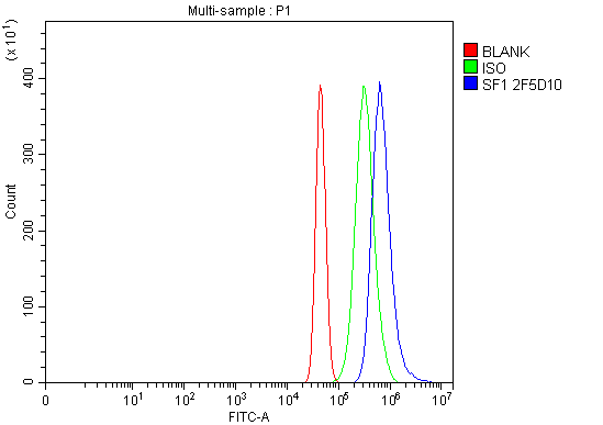
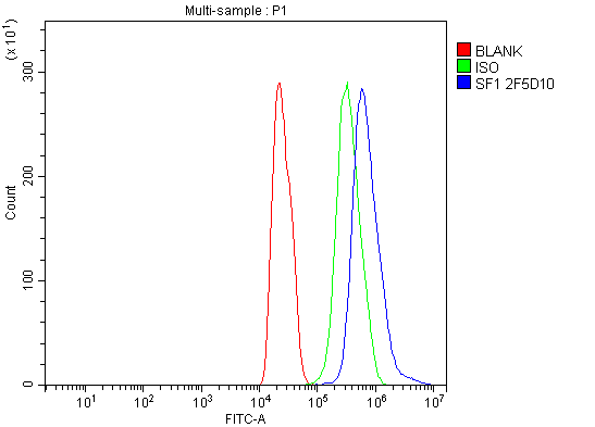
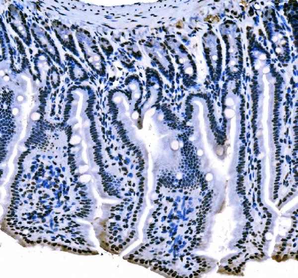
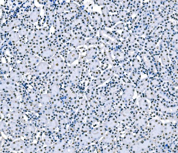
| WB, IHC, IF, ICC, FC |
|---|---|
| Primary Accession | Q15637 |
| Host | Mouse |
| Isotype | Mouse IgG2a |
| Reactivity | Rat, Human, Mouse |
| Clonality | Monoclonal |
| Format | Lyophilized |
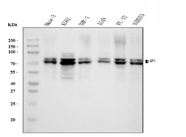
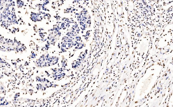
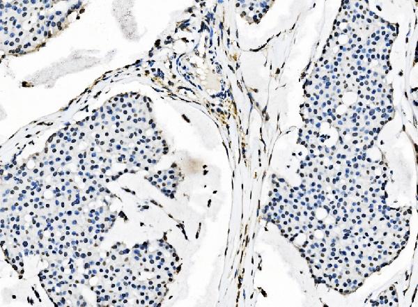
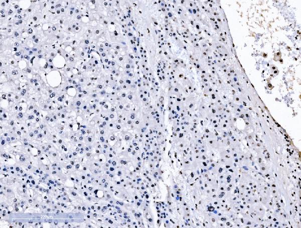
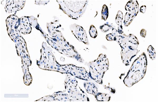
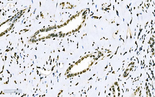
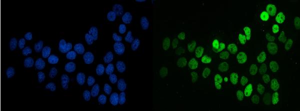
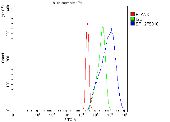
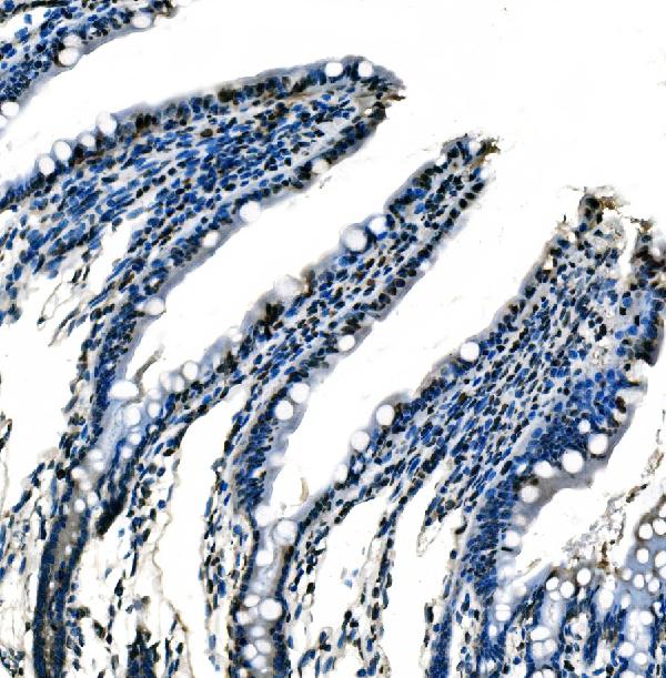
| Description | Anti-splicing factor 1 Antibody Picoband™ (monoclonal, 2F5D10) . Tested in Flow Cytometry, IF, IHC, ICC, WB applications. This antibody reacts with Human, Mouse, Rat. |
| Reconstitution | Adding 0.2 ml of distilled water will yield a concentration of 500 µg/ml. |
| Gene ID | 7536 |
|---|---|
| Other Names | Splicing factor 1, Mammalian branch point-binding protein, BBP, mBBP, Transcription factor ZFM1, Zinc finger gene in MEN1 locus, Zinc finger protein 162, SF1, ZFM1, ZNF162 |
| Calculated MW | 68 kDa |
| Application Details | Western blot, 0.25-0.5 µg/ml, Human, Mouse, Rat Immunohistochemistry(Paraffin-embedded Section), 2-5 µg/ml, Human, Mouse, Rat Immunocytochemistry/Immunofluorescence, 5 µg/ml, Human Flow Cytometry, 1-3 µg/1x10^6 cells, Human, Mouse, Rat |
| Contents | Each vial contains 4 mg Trehalose, 0.9 mg NaCl and 0.2 mg Na2HPO4. |
| Clone Names | Clone: 2F5D10 |
| Immunogen | E. coli-derived human splicing factor 1 recombinant protein (Position: R160-Q266). |
| Purification | Immunogen affinity purified. |
| Storage | At -20°C for one year from date of receipt. After reconstitution, at 4°C for one month. It can also be aliquotted and stored frozen at -20°C for six months. Avoid repeated freezing and thawing. |
| Name | SF1 |
|---|---|
| Synonyms | ZFM1, ZNF162 |
| Function | Necessary for the ATP-dependent first step of spliceosome assembly. Binds to the intron branch point sequence (BPS) 5'-UACUAAC-3' of the pre-mRNA. May act as transcription repressor. |
| Cellular Location | Nucleus. |
| Tissue Location | Detected in lung, ovary, adrenal gland, colon, kidney, muscle, pancreas, thyroid, placenta, brain, liver and heart |

Thousands of laboratories across the world have published research that depended on the performance of antibodies from Abcepta to advance their research. Check out links to articles that cite our products in major peer-reviewed journals, organized by research category.
info@abcepta.com, and receive a free "I Love Antibodies" mug.

Provided below are standard protocols that you may find useful for product applications.
Background
Splicing factor 1 also known as zinc finger protein 162 (ZFM162) is a protein that in humans is encoded by the SF1 gene. This gene encodes a nuclear pre-mRNA splicing factor. The encoded protein specifically recognizes the intron branch point sequence at the 3' splice site, together with the large subunit of U2 auxiliary factor (U2AF), and is required for the early stages of spliceosome assembly. It also plays a role in nuclear pre-mRNA retention and transcriptional repression. The encoded protein contains an N-terminal U2AF ligand motif, a central hnRNP K homology motif and quaking 2 region which bind a key branch-site adenosine within the branch point sequence, a zinc knuckles domain, and a C-terminal proline-rich domain. Alternative splicing results in multiple transcript variants.
If you have used an Abcepta product and would like to share how it has performed, please click on the "Submit Review" button and provide the requested information. Our staff will examine and post your review and contact you if needed.
If you have any additional inquiries please email technical services at tech@abcepta.com.
Ordering Information
Other Products
Shipping Information
