


 Foundational characteristics of cancer include proliferation, angiogenesis, migration, evasion of apoptosis, and cellular immortality. Find key markers for these cellular processes and antibodies to detect them.
 The SUMOplot™ Analysis Program predicts and scores sumoylation sites in your protein. SUMOylation is a post-translational modification involved in various cellular processes, such as nuclear-cytosolic transport, transcriptional regulation, apoptosis, protein stability, response to stress, and progression through the cell cycle.
 The Autophagy Receptor Motif Plotter predicts and scores autophagy receptor binding sites in your protein. Identifying proteins connected to this pathway is critical to understanding the role of autophagy in physiological as well as pathological processes such as development, differentiation, neurodegenerative diseases, stress, infection, and cancer.
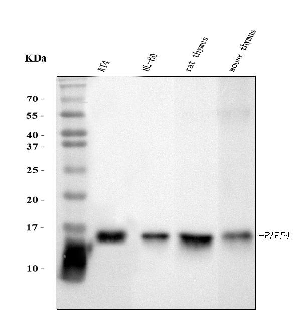
Anti-FABP4 Antibody Picoband™ (monoclonal, 10E12)
- SPECIFICATION
- CITATIONS
- PROTOCOLS
- BACKGROUND

Application
| WB, IHC |
|---|---|
| Primary Accession | P15090 |
| Host | Mouse |
| Isotype | Mouse IgG1 |
| Reactivity | Rat, Human, Mouse |
| Clonality | Monoclonal |
| Format | Lyophilized |
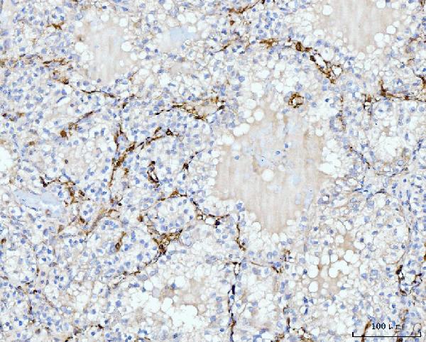
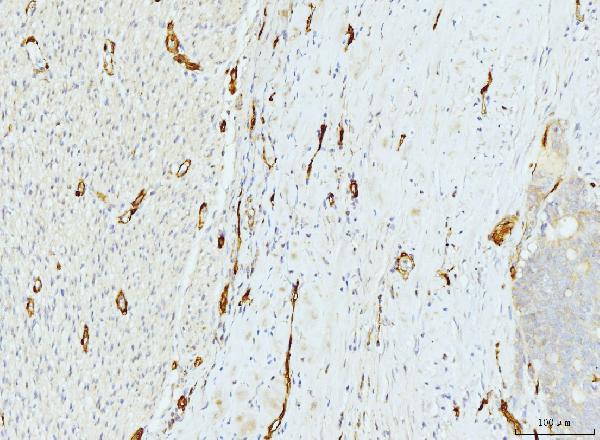
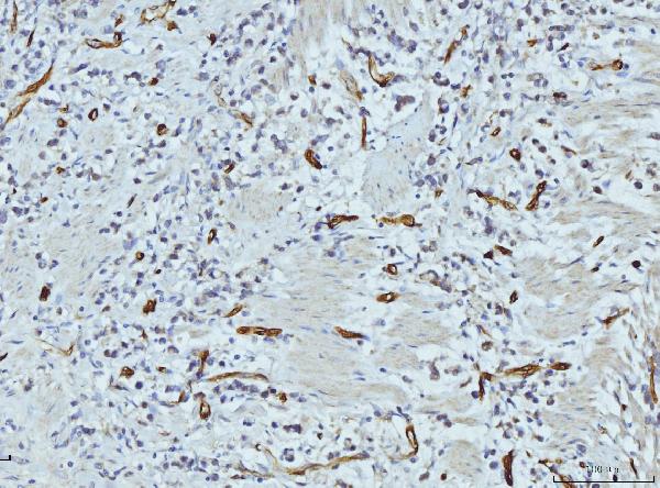
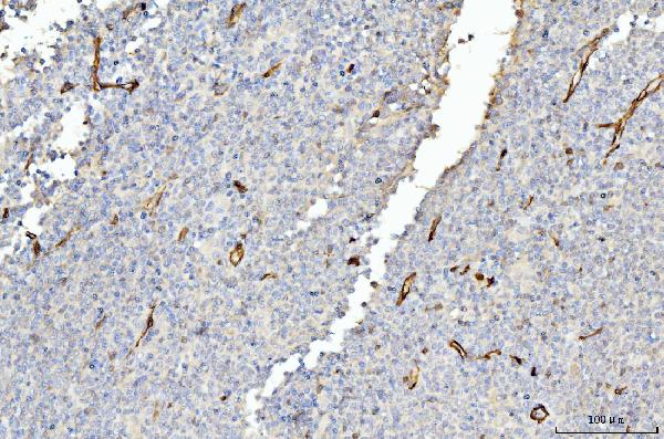
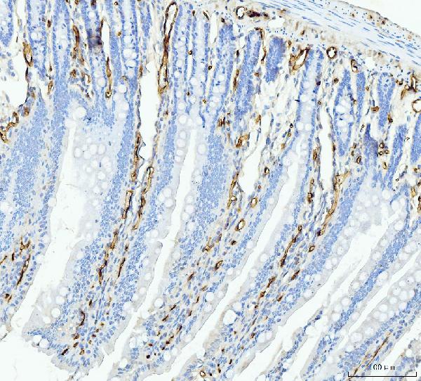
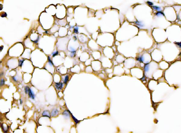
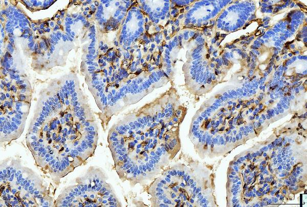
| Description | Anti-FABP4 Antibody Picoband™ (monoclonal, 10E12) . Tested in IHC, WB applications. This antibody reacts with Human, Mouse, Rat. |
| Reconstitution | Adding 0.2 ml of distilled water will yield a concentration of 500 µg/ml. |
| Gene ID | 2167 |
|---|---|
| Other Names | Fatty acid-binding protein, adipocyte, Adipocyte lipid-binding protein, ALBP, Adipocyte-type fatty acid-binding protein, A-FABP, AFABP, Fatty acid-binding protein 4, FABP4 |
| Calculated MW | 15 kDa |
| Application Details | Western blot, 0.25-0.5 µg/ml, Human, Mouse, Rat Immunohistochemistry(Paraffin-embedded Section), 2-5 µg/ml, Human, Mouse, Rat |
| Contents | Each vial contains 4 mg Trehalose, 0.9 mg NaCl and 0.2 mg Na2HPO4. |
| Clone Names | Clone: 10E12 |
| Immunogen | A synthetic peptide corresponding to a sequence at the N-terminus of human FABP4, identical to the related mouse and rat sequences. |
| Purification | Immunogen affinity purified. |
| Storage | At -20°C for one year from date of receipt. After reconstitution, at 4°C for one month. It can also be aliquotted and stored frozen at -20°C for six months. Avoid repeated freezing and thawing. |
| Name | FABP4 |
|---|---|
| Function | Lipid transport protein in adipocytes. Binds both long chain fatty acids and retinoic acid. Delivers long-chain fatty acids and retinoic acid to their cognate receptors in the nucleus. |
| Cellular Location | Cytoplasm {ECO:0000250|UniProtKB:P04117}. Nucleus {ECO:0000250|UniProtKB:P04117}. Note=Depending on the nature of the ligand, a conformation change exposes a nuclear localization motif and the protein is transported into the nucleus. Subject to constitutive nuclear export. {ECO:0000250|UniProtKB:P04117} |

Thousands of laboratories across the world have published research that depended on the performance of antibodies from Abcepta to advance their research. Check out links to articles that cite our products in major peer-reviewed journals, organized by research category.
info@abcepta.com, and receive a free "I Love Antibodies" mug.

Provided below are standard protocols that you may find useful for product applications.
Background
Fatty acid binding proteins (FABPs) are small cytoplasmic proteins that are expressed in a highly tissue-specific manner and bind to fatty acids such as oleic and retinoic acid. Adipocyte fatty-acid-binding protein, aP2 (FABP4) is expressed in adipocytes and macrophages, and integrates inflammatory and metabolic responses. Studies in aP2-deficient mice have shown that this lipid chaperone has a significant role in several aspects of metabolic syndrome, including type 2 diabetes and atherosclerosis. It regulates allergic airway inflammation and may provide a link between fatty acid metabolism and asthma.
If you have used an Abcepta product and would like to share how it has performed, please click on the "Submit Review" button and provide the requested information. Our staff will examine and post your review and contact you if needed.
If you have any additional inquiries please email technical services at tech@abcepta.com.
Ordering Information
Other Products
Shipping Information
