


 Foundational characteristics of cancer include proliferation, angiogenesis, migration, evasion of apoptosis, and cellular immortality. Find key markers for these cellular processes and antibodies to detect them.
 The SUMOplot™ Analysis Program predicts and scores sumoylation sites in your protein. SUMOylation is a post-translational modification involved in various cellular processes, such as nuclear-cytosolic transport, transcriptional regulation, apoptosis, protein stability, response to stress, and progression through the cell cycle.
 The Autophagy Receptor Motif Plotter predicts and scores autophagy receptor binding sites in your protein. Identifying proteins connected to this pathway is critical to understanding the role of autophagy in physiological as well as pathological processes such as development, differentiation, neurodegenerative diseases, stress, infection, and cancer.
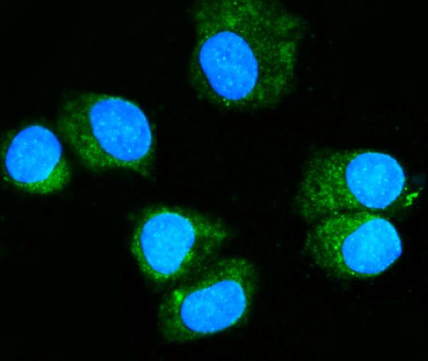
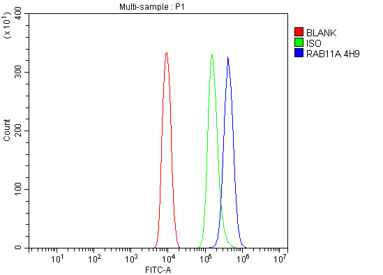
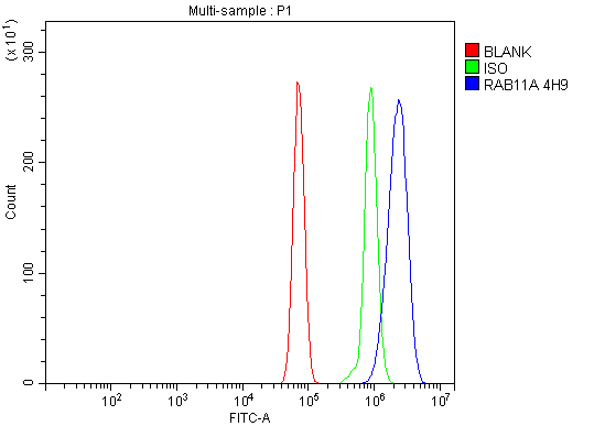
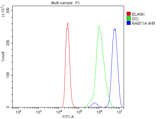
Anti-Rab11A Antibody Picoband™ (monoclonal, 4H9)
- SPECIFICATION
- CITATIONS
- PROTOCOLS
- BACKGROUND

Application
| WB, IHC, IF, ICC, FC |
|---|---|
| Primary Accession | P62491 |
| Host | Mouse |
| Isotype | Mouse IgG2b |
| Reactivity | Rat, Human, Mouse |
| Clonality | Monoclonal |
| Format | Lyophilized |
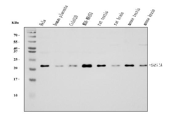
| Description | Anti-Rab11A Antibody Picoband™ (monoclonal, 4H9) . Tested in Flow Cytometry, IF, IHC, ICC, WB applications. This antibody reacts with Human, Mouse, Rat. |
| Reconstitution | Adding 0.2 ml of distilled water will yield a concentration of 500 µg/ml. |
| Gene ID | 8766 |
|---|---|
| Other Names | Ras-related protein Rab-11A, Rab-11, 3.6.5.2, YL8, RAB11A (HGNC:9760) |
| Calculated MW | 22 kDa |
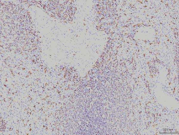
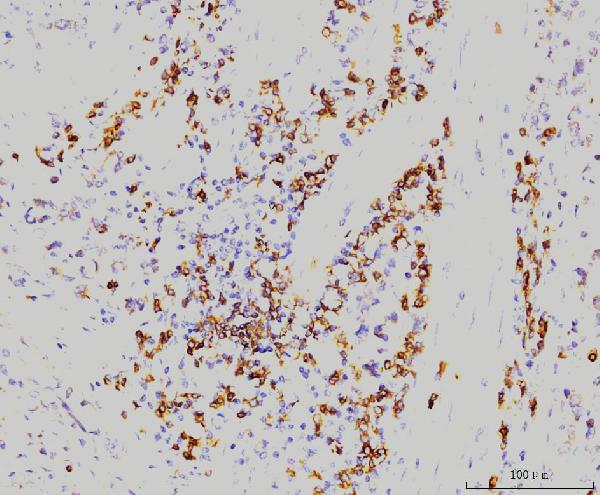
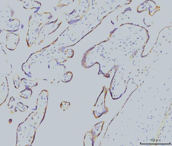
| Application Details | Western blot, 0.25-0.5 µg/ml, Human, Mouse, Rat Immunohistochemistry(Paraffin-embedded Section), 2-5 µg/ml, Human Immunocytochemistry/Immunofluorescence, 5 µg/ml, Human Flow Cytometry, 1-3 µg/1x10^6 cells, Human, Mouse, Rat |
| Contents | Each vial contains 4 mg Trehalose, 0.9 mg NaCl and 0.2 mg Na2HPO4. |
| Clone Names | Clone: 4H9 |
| Immunogen | A synthetic peptide corresponding to a sequence at the C-terminus of human Rab11A, identical to the related mouse and rat sequences. |
| Purification | Immunogen affinity purified. |
| Storage | At -20°C for one year from date of receipt. After reconstitution, at 4°C for one month. It can also be aliquotted and stored frozen at -20°C for six months. Avoid repeated freezing and thawing. |
| Name | RAB11A (HGNC:9760) |
|---|---|
| Function | The small GTPases Rab are key regulators of intracellular membrane trafficking, from the formation of transport vesicles to their fusion with membranes. Rabs cycle between an inactive GDP-bound form and an active GTP-bound form that is able to recruit to membranes different set of downstream effectors directly responsible for vesicle formation, movement, tethering and fusion (PubMed:15601896, PubMed:15689490, PubMed:17462998, PubMed:19542231, PubMed:20026645, PubMed:20890297, PubMed:21282656). The small Rab GTPase RAB11A regulates endocytic recycling (PubMed:20026645). Forms a functional Rab11/FIP3/dynein complex that regulates the movement of peripheral sorting endosomes (SE) along microtubule tracks toward the microtubule organizing center/centrosome, generating the endosomal recycling compartment (ERC) (PubMed:20026645). Acts as a major regulator of membrane delivery during cytokinesis (PubMed:15601896). Together with MYO5B and RAB8A participates in epithelial cell polarization. Together with RAB3IP, RAB8A, the exocyst complex, PARD3, PRKCI, ANXA2, CDC42 and DNMBP promotes transcytosis of PODXL to the apical membrane initiation sites (AMIS), apical surface formation and lumenogenesis. Together with MYO5B participates in CFTR trafficking to the plasma membrane and TF (Transferrin) recycling in nonpolarized cells. Required in a complex with MYO5B and RAB11FIP2 for the transport of NPC1L1 to the plasma membrane. Participates in the sorting and basolateral transport of CDH1 from the Golgi apparatus to the plasma membrane. Regulates the recycling of FCGRT (receptor of Fc region of monomeric Ig G) to basolateral membranes. May also play a role in melanosome transport and release from melanocytes (PubMed:15689490, PubMed:17462998, PubMed:19542231, PubMed:20890297, PubMed:21282656). Promotes Rabin8/RAB3IP preciliary vesicular trafficking to mother centriole by forming a ciliary targeting complex containing Rab11, ASAP1, Rabin8/RAB3IP, RAB11FIP3 and ARF4, thereby regulating ciliogenesis initiation (PubMed:25673879, PubMed:31204173). On the contrary, upon LPAR1 receptor signaling pathway activation, interaction with phosphorylated WDR44 prevents Rab11-RAB3IP-RAB11FIP3 complex formation and cilia growth (PubMed:31204173). Participates in the export of a subset of neosynthesized proteins through a Rab8-Rab10-Rab11- endososomal dependent export route via interaction with WDR44 (PubMed:32344433). |
| Cellular Location | Cell membrane; Lipid-anchor. Endosome membrane. Recycling endosome membrane; Lipid-anchor. Cleavage furrow. Cytoplasmic vesicle, phagosome. Cytoplasmic vesicle membrane. Golgi apparatus. Golgi apparatus, trans-Golgi network. Note=Localized to WDR44-positive endosomes and tubules (PubMed:32344433). Translocates with RAB11FIP2 from the vesicles of the endocytic recycling compartment (ERC) to the plasma membrane (PubMed:11994279). During interphase, localized in vesicles continuously moving from peripheral sorting endosomes towards the pericentrosomal ERC (PubMed:20026645). Localizes to the cleavage furrow (PubMed:15601896). Colocalizes with PARD3, PRKCI, EXOC5, OCLN, PODXL and RAB8A in apical membrane initiation sites (AMIS) during the generation of apical surface and lumenogenesis (PubMed:20890297) Recruited to phagosomes containing S.aureus or M.tuberculosis (PubMed:21255211). Localized to rhodopsin transport carriers when interacting with RAB11AFIP3 and ASAP1 in photoreceptors (PubMed:25673879). |

Thousands of laboratories across the world have published research that depended on the performance of antibodies from Abcepta to advance their research. Check out links to articles that cite our products in major peer-reviewed journals, organized by research category.
info@abcepta.com, and receive a free "I Love Antibodies" mug.

Provided below are standard protocols that you may find useful for product applications.
Background
Ras-related protein Rab-11A is a protein that in humans is encoded by the RAB11A gene. The protein encoded by this gene belongs to the small GTPase superfamily, Rab family which plays essential roles in vesicle and granule targeting. It is mapped to 15q22.31. RAB11A is associated with both constitutive and regulated secretory pathways, and may be involved in protein transport. Additionally, RAB11A can control intracellular trafficking of the innate immune receptor TLR4, and thereby also receptor signaling. It has been shown to interact with RAB11FIP2, RAB11FIP4, and RAB11FIP1 and so on.
If you have used an Abcepta product and would like to share how it has performed, please click on the "Submit Review" button and provide the requested information. Our staff will examine and post your review and contact you if needed.
If you have any additional inquiries please email technical services at tech@abcepta.com.
Ordering Information
Other Products
Shipping Information
