


 Foundational characteristics of cancer include proliferation, angiogenesis, migration, evasion of apoptosis, and cellular immortality. Find key markers for these cellular processes and antibodies to detect them.
 The SUMOplot™ Analysis Program predicts and scores sumoylation sites in your protein. SUMOylation is a post-translational modification involved in various cellular processes, such as nuclear-cytosolic transport, transcriptional regulation, apoptosis, protein stability, response to stress, and progression through the cell cycle.
 The Autophagy Receptor Motif Plotter predicts and scores autophagy receptor binding sites in your protein. Identifying proteins connected to this pathway is critical to understanding the role of autophagy in physiological as well as pathological processes such as development, differentiation, neurodegenerative diseases, stress, infection, and cancer.
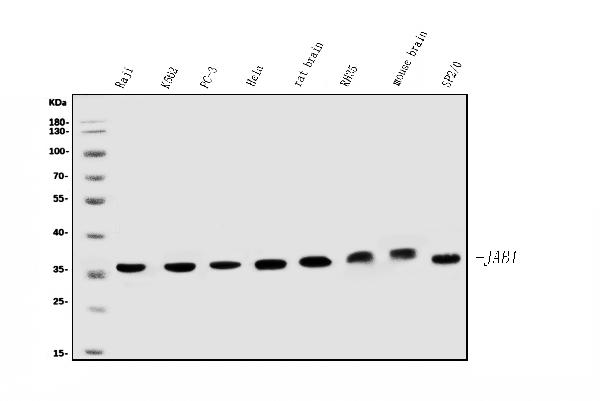
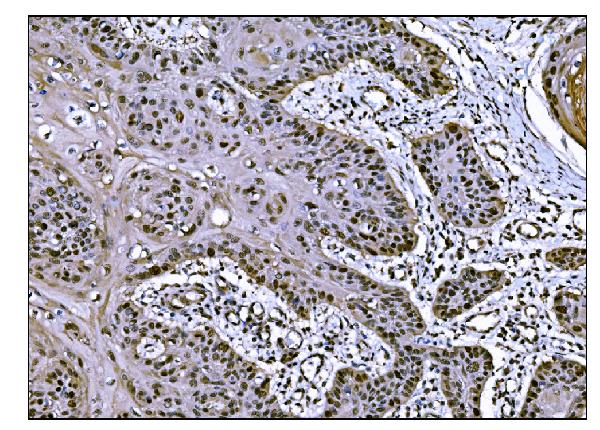
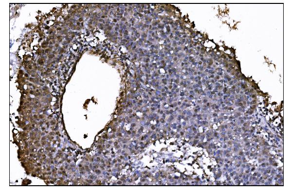
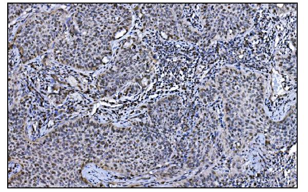
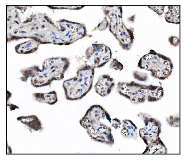
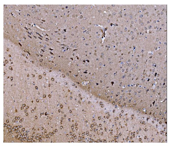
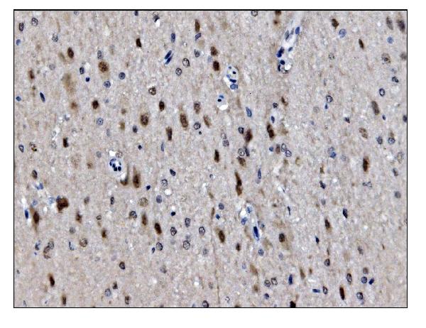
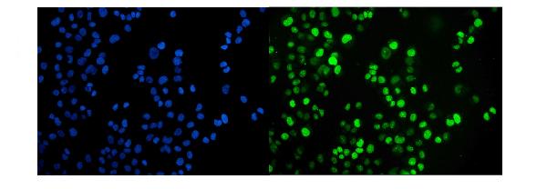
Anti-JAB1 Antibody Picoband™ (monoclonal, 4G9)
- SPECIFICATION
- CITATIONS
- PROTOCOLS
- BACKGROUND

Application
| WB, IHC, IF, ICC, FC |
|---|---|
| Primary Accession | Q92905 |
| Host | Mouse |
| Isotype | Mouse IgG2b |
| Reactivity | Rat, Human, Mouse |
| Clonality | Monoclonal |
| Format | Lyophilized |
| Description | Anti-JAB1 Antibody Picoband™ (monoclonal, 4G9) . Tested in Flow Cytometry, IF, IHC, ICC, WB applications. This antibody reacts with Human, Mouse, Rat. |
| Reconstitution | Add 0.2ml of distilled water will yield a concentration of 500ug/ml. |
| Gene ID | 10987 |
|---|---|
| Other Names | COP9 signalosome complex subunit 5, SGN5, Signalosome subunit 5, 3.4.-.-, Jun activation domain-binding protein 1, COPS5, CSN5, JAB1 |
| Calculated MW | 37 kDa |
| Application Details | Western blot, 0.25-0.5 µg/ml, Human, Mouse, Rat Immunohistochemistry (Paraffin-embedded Section), 2-5 µg/ml, Human, Mouse, Rat Immunocytochemistry/Immunofluorescence, 5 µg/ml, Human Flow Cytometry, 1-3 µg/1x10^6 cells, Human |
| Contents | Each vial contains 4mg Trehalose, 0.9mg NaCl and 0.2mg Na2HPO4. |
| Clone Names | Clone: 4G9 |
| Immunogen | E.coli-derived human JAB1 recombinant protein (Position: M8-S334). Human JAB1 shares 99.4% amino acid (aa) sequence identity with mouse JAB1. |
| Purification | Immunogen affinity purified. |
| Storage | Store at -20˚C for one year from date of receipt. After reconstitution, at 4˚C for one month. It can also be aliquotted and stored frozen at -20˚C for six months. Avoid repeated freeze-thaw cycles. |
| Name | COPS5 |
|---|---|
| Synonyms | CSN5, JAB1 |
| Function | Probable protease subunit of the COP9 signalosome complex (CSN), a complex involved in various cellular and developmental processes. The CSN complex is an essential regulator of the ubiquitin (Ubl) conjugation pathway by mediating the deneddylation of the cullin subunits of the SCF-type E3 ligase complexes, leading to decrease the Ubl ligase activity of SCF-type complexes such as SCF, CSA or DDB2. The complex is also involved in phosphorylation of p53/TP53, c-jun/JUN, IkappaBalpha/NFKBIA, ITPK1 and IRF8, possibly via its association with CK2 and PKD kinases. CSN-dependent phosphorylation of TP53 and JUN promotes and protects degradation by the Ubl system, respectively. In the complex, it probably acts as the catalytic center that mediates the cleavage of Nedd8 from cullins. It however has no metalloprotease activity by itself and requires the other subunits of the CSN complex. Interacts directly with a large number of proteins that are regulated by the CSN complex, confirming a key role in the complex. Promotes the proteasomal degradation of BRSK2. |
| Cellular Location | Cytoplasm, cytosol. Nucleus. Cytoplasm, perinuclear region. Cytoplasmic vesicle, secretory vesicle, synaptic vesicle Note=Nuclear localization is diminished in the presence of IFIT3 |

Thousands of laboratories across the world have published research that depended on the performance of antibodies from Abcepta to advance their research. Check out links to articles that cite our products in major peer-reviewed journals, organized by research category.
info@abcepta.com, and receive a free "I Love Antibodies" mug.

Provided below are standard protocols that you may find useful for product applications.
Background
COP9 constitutive photomorphogenic homolog subunit 5 (Arabidopsis), also known as COPS5 or JAB1, is a gene conserved from humans to Saccharomyces cerevisiae. It is a member of the MOV34 family. COPS5 is mapped to 8q13.1. The protein encoded by this gene is one of the eight subunits of COP9 signalosome, a highly conserved protein complex that functions as an important regulator in multiple signaling pathways. COPS5 can interact with the cytoplasmic domain of the beta-2 subunit of the alpha-L/beta-2 integrin LFA1, and it is the only protein demonstrated to interact with MIF. COPS5, VHL, and TRC8 proteins appear to be linked both physically and functionally, and all 3 may participate in the development of kidney cancer. In addition to that, COPS5 is an essential cofactor for the apoptotic function of E2F1.
If you have used an Abcepta product and would like to share how it has performed, please click on the "Submit Review" button and provide the requested information. Our staff will examine and post your review and contact you if needed.
If you have any additional inquiries please email technical services at tech@abcepta.com.
Ordering Information
Other Products
Shipping Information
