


 Foundational characteristics of cancer include proliferation, angiogenesis, migration, evasion of apoptosis, and cellular immortality. Find key markers for these cellular processes and antibodies to detect them.
 The SUMOplot™ Analysis Program predicts and scores sumoylation sites in your protein. SUMOylation is a post-translational modification involved in various cellular processes, such as nuclear-cytosolic transport, transcriptional regulation, apoptosis, protein stability, response to stress, and progression through the cell cycle.
 The Autophagy Receptor Motif Plotter predicts and scores autophagy receptor binding sites in your protein. Identifying proteins connected to this pathway is critical to understanding the role of autophagy in physiological as well as pathological processes such as development, differentiation, neurodegenerative diseases, stress, infection, and cancer.
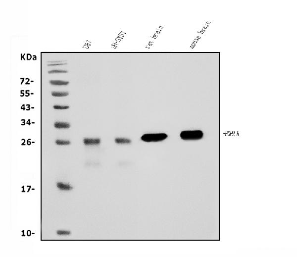
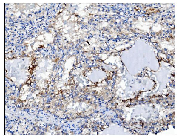
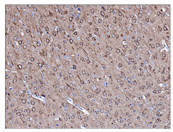
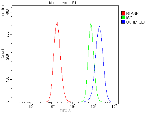
Anti-PGP9.5 Antibody Picoband™ (monoclonal, 3E4)
- SPECIFICATION
- CITATIONS
- PROTOCOLS
- BACKGROUND

Application
| WB, IHC, FC |
|---|---|
| Primary Accession | P09936 |
| Host | Mouse |
| Isotype | Mouse IgG1 |
| Reactivity | Rat, Human, Mouse |
| Clonality | Monoclonal |
| Format | Lyophilized |
| Description | Anti-PGP9.5 Antibody Picoband™ (monoclonal, 3E4) . Tested in Flow Cytometry, IHC, WB applications. This antibody reacts with Human, Mouse, Rat. |
| Reconstitution | Add 0.2ml of distilled water will yield a concentration of 500ug/ml. |
| Gene ID | 7345 |
|---|---|
| Other Names | Ubiquitin carboxyl-terminal hydrolase isozyme L1, UCH-L1, 3.4.19.12, Neuron cytoplasmic protein 9.5, PGP 9.5, PGP9.5, Ubiquitin thioesterase L1, UCHL1 |
| Calculated MW | 27 kDa |
| Application Details | Western blot, 0.25-0.5 µg/ml, Human, Mouse, Rat Immunohistochemistry (Paraffin-embedded Section), 2-5 µg/ml, Human, Rat Flow Cytometry, 1-3 µg/1x10^6 cells, Human |
| Contents | Each vial contains 4mg Trehalose, 0.9mg NaCl and 0.2mg Na2HPO4. |
| Clone Names | Clone: 3E4 |
| Immunogen | A synthetic peptide corresponding to a sequence at the C-terminus of human PGP9.5, different from the related mouse and rat sequences by two amino acids. |
| Purification | Immunogen affinity purified. |
| Storage | Store at -20˚C for one year from date of receipt. After reconstitution, at 4˚C for one month. It can also be aliquotted and stored frozen at -20˚C for six months. Avoid repeated freeze-thaw cycles. |
| Name | UCHL1 |
|---|---|
| Function | Deubiquitinase that plays a role in the regulation of several processes such as maintenance of synaptic function, cardiac function, inflammatory response or osteoclastogenesis (PubMed:22212137, PubMed:23359680). Abrogates the ubiquitination of multiple proteins including WWTR1/TAZ, EGFR, HIF1A and beta-site amyloid precursor protein cleaving enzyme 1/BACE1 (PubMed:22212137, PubMed:25615526). In addition, recognizes and hydrolyzes a peptide bond at the C-terminal glycine of ubiquitin to maintain a stable pool of monoubiquitin that is a key requirement for the ubiquitin-proteasome and the autophagy- lysosome pathways (PubMed:12408865, PubMed:8639624, PubMed:9774100). Regulates amyloid precursor protein/APP processing by promoting BACE1 degradation resulting in decreased amyloid beta production (PubMed:22212137). Plays a role in the immune response by regulating the ability of MHC I molecules to reach cross-presentation compartments competent for generating Ag-MHC I complexes (By similarity). Mediates the 'Lys-48'-linked deubiquitination of the transcriptional coactivator WWTR1/TAZ leading to its stabilization and inhibition of osteoclastogenesis (By similarity). Deubiquitinates and stabilizes epidermal growth factor receptor EGFR to prevent its degradation and to activate its downstream mediators (By similarity). Modulates oxidative activity in skeletal muscle by regulating key mitochondrial oxidative proteins (By similarity). Enhances the activity of hypoxia-inducible factor 1-alpha/HIF1A by abrogateing its VHL E3 ligase-mediated ubiquitination and consequently inhibiting its degradation (PubMed:25615526). |
| Cellular Location | Cytoplasm. Endoplasmic reticulum membrane; Lipid- anchor. Note=About 30% of total UCHL1 is associated with membranes in brain. Localizes near and/or within mitochondria to potentially interact with mitochondrial proteins {ECO:0000250|UniProtKB:Q9R0P9} |
| Tissue Location | Found in neuronal cell bodies and processes throughout the neocortex (at protein level). Expressed in neurons and cells of the diffuse neuroendocrine system and their tumors. Weakly expressed in ovary. Down-regulated in brains from Parkinson disease and Alzheimer disease patients. |

Thousands of laboratories across the world have published research that depended on the performance of antibodies from Abcepta to advance their research. Check out links to articles that cite our products in major peer-reviewed journals, organized by research category.
info@abcepta.com, and receive a free "I Love Antibodies" mug.

Provided below are standard protocols that you may find useful for product applications.
Background
UCH-L1, also known as PGP9.5, is a member of a gene family whose products hydrolyze small C-terminal adducts of ubiquitin to generate the ubiquitin monomer. Expression of UCH-L1 is highly specific to neurons and to cells of the diffuse neuroendocrine system and their tumors. It is abundantly present in all neurons (accounts for 1-2% of total brain protein), expressed specifically in neurons and testis/ovary. The catalytic triad of UCH-L1 contains a cysteine at position 90, an aspartate at position 176, and a histidine at position 161 that are responsible for its hydrolase activity.
If you have used an Abcepta product and would like to share how it has performed, please click on the "Submit Review" button and provide the requested information. Our staff will examine and post your review and contact you if needed.
If you have any additional inquiries please email technical services at tech@abcepta.com.
Ordering Information
Other Products
Shipping Information
