


 Foundational characteristics of cancer include proliferation, angiogenesis, migration, evasion of apoptosis, and cellular immortality. Find key markers for these cellular processes and antibodies to detect them.
 The SUMOplot™ Analysis Program predicts and scores sumoylation sites in your protein. SUMOylation is a post-translational modification involved in various cellular processes, such as nuclear-cytosolic transport, transcriptional regulation, apoptosis, protein stability, response to stress, and progression through the cell cycle.
 The Autophagy Receptor Motif Plotter predicts and scores autophagy receptor binding sites in your protein. Identifying proteins connected to this pathway is critical to understanding the role of autophagy in physiological as well as pathological processes such as development, differentiation, neurodegenerative diseases, stress, infection, and cancer.
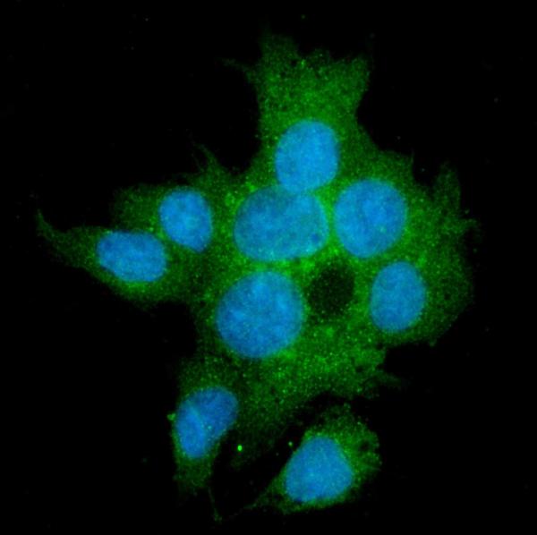
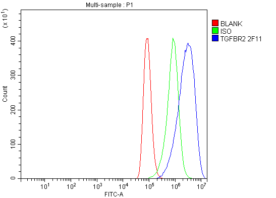
Anti-TGFBR2 Antibody Picoband™ (monoclonal, 2F11)
- SPECIFICATION
- CITATIONS
- PROTOCOLS
- BACKGROUND

Application
| WB, IHC, IF, ICC, FC |
|---|---|
| Primary Accession | P37173 |
| Host | Mouse |
| Isotype | Mouse IgG2b |
| Reactivity | Human |
| Clonality | Monoclonal |
| Format | Lyophilized |
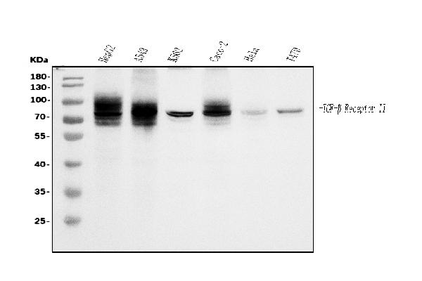
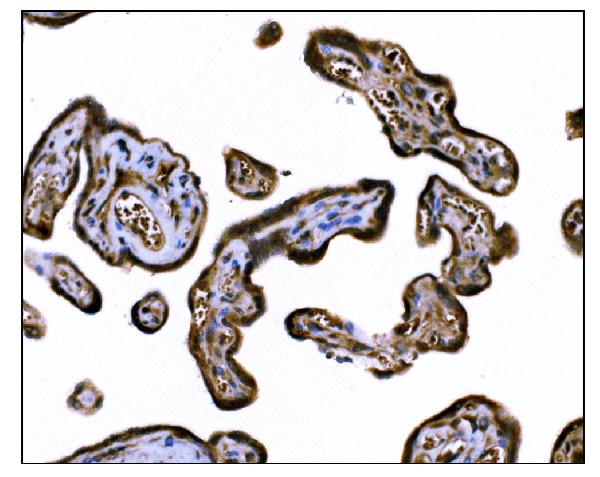
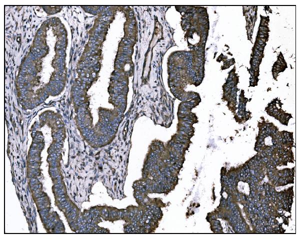
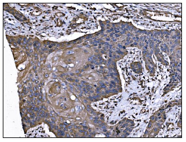
| Description | Anti-TGFBR2 Antibody Picoband™ (monoclonal, 2F11) . Tested in Flow Cytometry, IF, IHC, ICC, WB applications. This antibody reacts with Human. |
| Reconstitution | Add 0.2ml of distilled water will yield a concentration of 500ug/ml. |
| Gene ID | 7048 |
|---|---|
| Other Names | TGF-beta receptor type-2, TGFR-2, 2.7.11.30, TGF-beta type II receptor, Transforming growth factor-beta receptor type II, TGF-beta receptor type II, TbetaR-II, TGFBR2 |
| Calculated MW | 70-85 kDa |
| Application Details | Western blot, 0.25-0.5 µg/ml, Human Immunohistochemistry (Paraffin-embedded Section), 2-5 µg/ml, Human Immunocytochemistry/Immunofluorescence, 5 µg/ml, Human Flow Cytometry, 1-3 µg/1x10^6 cells, Human |
| Contents | Each vial contains 4mg Trehalose, 0.9mg NaCl and 0.2mg Na2HPO4. |
| Clone Names | Clone: 2F11 |
| Immunogen | A synthetic peptide corresponding to a sequence at the N-terminus of human TGFBR2, different from the related mouse sequence by five amino acids, and from the related rat sequence by eight amino acids. |
| Purification | Immunogen affinity purified. |
| Storage | Store at -20˚C for one year from date of receipt. After reconstitution, at 4˚C for one month. It can also be aliquotted and stored frozen at -20˚C for six months. Avoid repeated freeze-thaw cycles. |
| Name | TGFBR2 |
|---|---|
| Function | Transmembrane serine/threonine kinase forming with the TGF- beta type I serine/threonine kinase receptor, TGFBR1, the non- promiscuous receptor for the TGF-beta cytokines TGFB1, TGFB2 and TGFB3. Transduces the TGFB1, TGFB2 and TGFB3 signal from the cell surface to the cytoplasm and thus regulates a plethora of physiological and pathological processes including cell cycle arrest in epithelial and hematopoietic cells, control of mesenchymal cell proliferation and differentiation, wound healing, extracellular matrix production, immunosuppression and carcinogenesis. The formation of the receptor complex composed of 2 TGFBR1 and 2 TGFBR2 molecules symmetrically bound to the cytokine dimer results in the phosphorylation and activation of TGFBR1 by the constitutively active TGFBR2. Activated TGFBR1 phosphorylates SMAD2 which dissociates from the receptor and interacts with SMAD4. The SMAD2-SMAD4 complex is subsequently translocated to the nucleus where it modulates the transcription of the TGF-beta-regulated genes. This constitutes the canonical SMAD-dependent TGF-beta signaling cascade. Also involved in non-canonical, SMAD-independent TGF-beta signaling pathways. |
| Cellular Location | Cell membrane; Single-pass type I membrane protein. Membrane raft |

Thousands of laboratories across the world have published research that depended on the performance of antibodies from Abcepta to advance their research. Check out links to articles that cite our products in major peer-reviewed journals, organized by research category.
info@abcepta.com, and receive a free "I Love Antibodies" mug.

Provided below are standard protocols that you may find useful for product applications.
Background
TGFBR2 (transforming growth factor, beta receptor II (70/80kDa)), also known as TGF-beta receptor type-2, TGFR-2, TGF-beta type II receptor, Transforming growth factor-beta receptor type II (TGF-beta receptor type II, TbetaR-II), is a member of the Ser/Thr protein kinase family and the TGFB receptor subfamily. A TGFBR2 cDNA encodes a deduced 565-amino acid protein with a calculated molecular mass of approximately 60 kD in length. The encoded protein is a transmembrane protein that has a protein kinase domain, forms a heterodimeric complex with another receptor protein, and binds TGF-beta. This receptor/ligand complex phosphorylates proteins, which then enter the nucleus and regulate the transcription of a subset of genes related to cell proliferation. Mutations in this gene have been associated with Marfan syndrome, Loeys-Deitz aortic aneurysm syndrome, Osler-Weber-Rendu syndrome, and the development of various types of tumors. Alternatively spliced transcript variants encoding different informs have been characterized.
If you have used an Abcepta product and would like to share how it has performed, please click on the "Submit Review" button and provide the requested information. Our staff will examine and post your review and contact you if needed.
If you have any additional inquiries please email technical services at tech@abcepta.com.
Ordering Information
Other Products
Shipping Information
