


 Foundational characteristics of cancer include proliferation, angiogenesis, migration, evasion of apoptosis, and cellular immortality. Find key markers for these cellular processes and antibodies to detect them.
 The SUMOplot™ Analysis Program predicts and scores sumoylation sites in your protein. SUMOylation is a post-translational modification involved in various cellular processes, such as nuclear-cytosolic transport, transcriptional regulation, apoptosis, protein stability, response to stress, and progression through the cell cycle.
 The Autophagy Receptor Motif Plotter predicts and scores autophagy receptor binding sites in your protein. Identifying proteins connected to this pathway is critical to understanding the role of autophagy in physiological as well as pathological processes such as development, differentiation, neurodegenerative diseases, stress, infection, and cancer.
Anti-Ki67 Antibody Picoband™ (monoclonal, 5E12)
- SPECIFICATION
- CITATIONS
- PROTOCOLS
- BACKGROUND

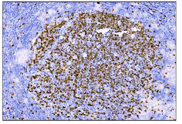
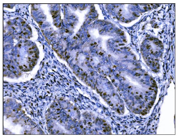
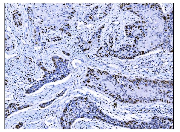
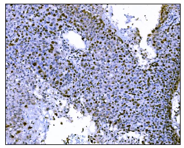
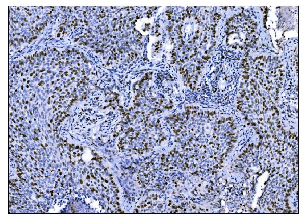
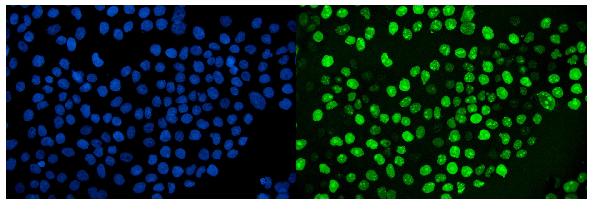
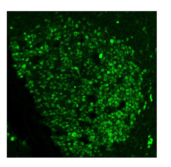
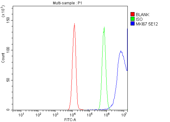
Application
| IHC, IF, ICC, FC |
|---|---|
| Primary Accession | P46013 |
| Host | Mouse |
| Isotype | Mouse IgG2b |
| Reactivity | Human |
| Clonality | Monoclonal |
| Format | Lyophilized |
| Description | Anti-Ki67 Antibody Picoband™ (monoclonal, 5E12) . Tested in Flow Cytometry, IF, IHC, ICC applications. This antibody reacts with Human. |
| Reconstitution | Add 0.2ml of distilled water will yield a concentration of 500ug/ml. |
| Gene ID | 4288 |
|---|---|
| Other Names | Proliferation marker protein Ki-67, Antigen identified by monoclonal antibody Ki-67, Antigen KI-67, Antigen Ki67, MKI67 (HGNC:7107) |
| Calculated MW | 358694 Da |
| Application Details | Immunohistochemistry (Paraffin-embedded Section), 2-5 µg/ml, Human Immunocytochemistry/Immunofluorescence, 5 µg/ml, Human Immunofluorescence, 5 µg/ml, Human Flow Cytometry, 1-3 µg/1x10^6 cells, Human |
| Contents | Each vial contains 4mg Trehalose, 0.9mg NaCl and 0.2mg Na2HPO4. |
| Clone Names | Clone: 5E12 |
| Immunogen | E. coli-derived human Ki67 recombinant protein (Position: K2860-I3256). |
| Purification | Immunogen affinity purified. |
| Storage | Store at -20˚C for one year from date of receipt. After reconstitution, at 4˚C for one month. It can also be aliquotted and stored frozen at -20˚C for six months. Avoid repeated freeze-thaw cycles. |
| Name | MKI67 (HGNC:7107) |
|---|---|
| Function | Protein that associates with the surface of mitotic chromosomes and acts both as a chromosome repellent during early mitosis and chromosome attractant during late mitosis (PubMed:27362226, PubMed:32879492, PubMed:35513709, PubMed:39153474). Required to maintain individual mitotic chromosomes dispersed in the cytoplasm following nuclear envelope disassembly (PubMed:27362226). During early mitosis, relocalizes from nucleoli to the chromosome surface where it forms extended brush structures that cover a substantial fraction of the chromosome surface (PubMed:27362226). The MKI67 brush structure prevents chromosomes from collapsing into a single chromatin mass by forming a steric and electrostatic charge barrier: the protein has a high net electrical charge and acts as a surfactant, dispersing chromosomes and enabling independent chromosome motility (PubMed:27362226). During mitotic anaphase, the MKI67 brush structure collapses and MKI67 switches from a chromosome repellent to a chromosome attractant to promote chromosome clustering and facilitate the exclusion of large cytoplasmic particles from the future nuclear space (PubMed:32879492, PubMed:39153474). Mechanistically, dephosphorylation during mitotic exit and simultaneous exposure of a conserved basic patch induce the RNA-dependent formation of a liquid- like condensed phase on the chromosome surface, promoting coalescence of neighboring chromosome surfaces and clustering of chromosomes (PubMed:39153474). Binds premature ribosomal RNAs during anaphase; promoting liquid-liquid phase separation (PubMed:28935370, PubMed:39153474). Binds DNA, with a preference for supercoiled DNA and AT-rich DNA (PubMed:10878551). Does not contribute to the internal structure of mitotic chromosomes (By similarity). May play a role in chromatin organization; it is however unclear whether it plays a direct role in chromatin organization or whether it is an indirect consequence of its function in mitotic chromosome (PubMed:24867636). |
| Cellular Location | Chromosome. Nucleus. Nucleus, nucleolus. Note=During early mitosis, relocalizes from nucleoli to the surface of the mitotic chromosome, the perichromosomal layer, and covers a substantial fraction of the mitotic chromosome surface (PubMed:27362226) Associates with satellite DNA in G1 phase (PubMed:9510506). Binds tightly to chromatin in interphase, chromatin-binding decreases in mitosis when it associates with the surface of the condensed chromosomes (PubMed:15896774, PubMed:22002106). Predominantly localized in the G1 phase in the perinucleolar region, in the later phases it is also detected throughout the nuclear interior, being predominantly localized in the nuclear matrix (PubMed:22002106) |

Thousands of laboratories across the world have published research that depended on the performance of antibodies from Abcepta to advance their research. Check out links to articles that cite our products in major peer-reviewed journals, organized by research category.
info@abcepta.com, and receive a free "I Love Antibodies" mug.

Provided below are standard protocols that you may find useful for product applications.
Background
Ki-67 (Proliferation-related Ki-67 antigen), also known as MKI67 or KIA, is a protein that in humans is encoded by the MKI67 gene. From study of a panel of human-rodent somatic cell hybrids, it has been demonstrated that a gene involved in the expression of the MKI67 antigen is located on chromosome 10. By in situ hybridization, Fonatsch et al. (1991) regionalized the MKI67 gene to chromosome 10q25-qter. By FISH, Traut et al. (1998) mapped the mouse Mki67 gene to chromosome 7F3-F5. Antigen KI-67 is a nuclear protein that is associated with and may be necessary for cellular proliferation. Furthermore it is associated with ribosomal RNA transcription. Inactivation of antigen KI-67 leads to inhibition of ribosomal RNA synthesis.
If you have used an Abcepta product and would like to share how it has performed, please click on the "Submit Review" button and provide the requested information. Our staff will examine and post your review and contact you if needed.
If you have any additional inquiries please email technical services at tech@abcepta.com.
Ordering Information
Other Products
Shipping Information
