


 Foundational characteristics of cancer include proliferation, angiogenesis, migration, evasion of apoptosis, and cellular immortality. Find key markers for these cellular processes and antibodies to detect them.
 The SUMOplot™ Analysis Program predicts and scores sumoylation sites in your protein. SUMOylation is a post-translational modification involved in various cellular processes, such as nuclear-cytosolic transport, transcriptional regulation, apoptosis, protein stability, response to stress, and progression through the cell cycle.
 The Autophagy Receptor Motif Plotter predicts and scores autophagy receptor binding sites in your protein. Identifying proteins connected to this pathway is critical to understanding the role of autophagy in physiological as well as pathological processes such as development, differentiation, neurodegenerative diseases, stress, infection, and cancer.
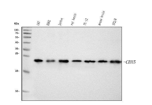
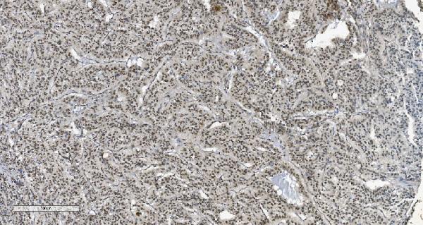
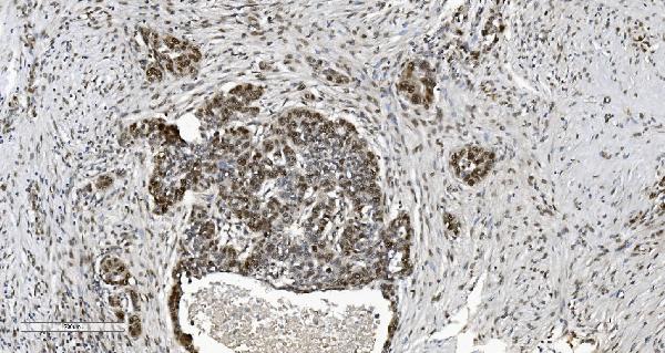
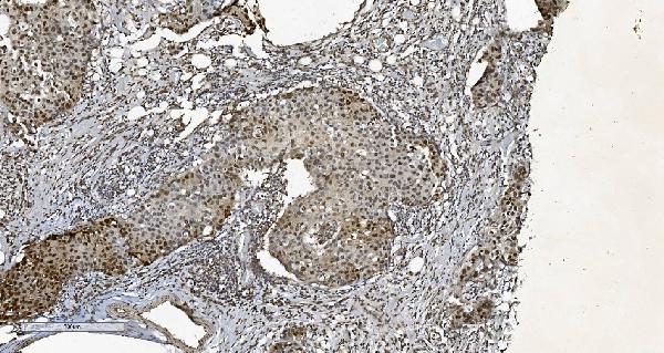
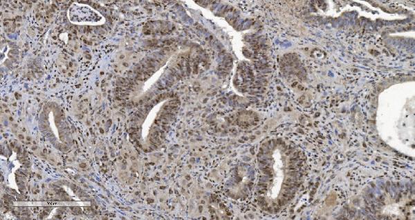
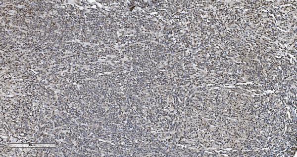
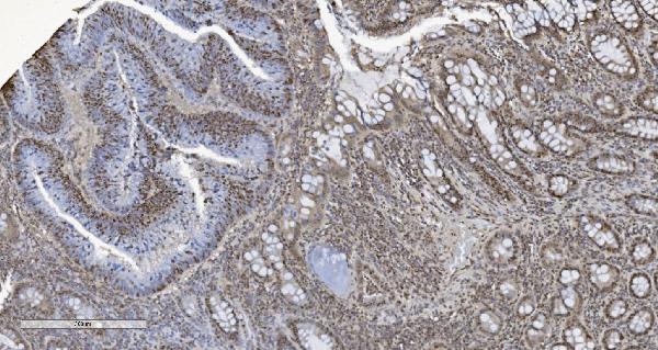
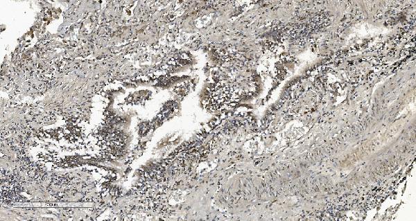
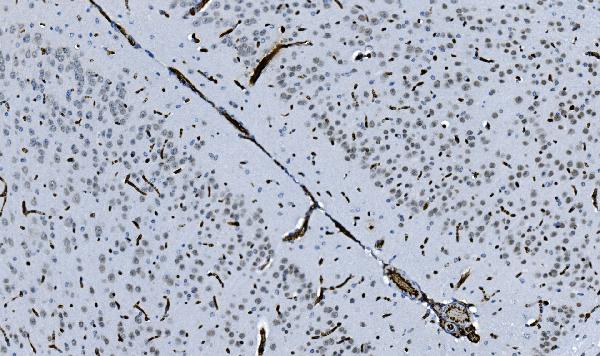
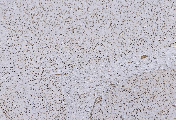
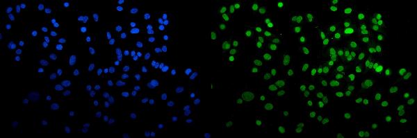
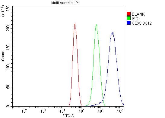
Anti-HP1 alpha/CBX5 Antibody Picoband™ (monoclonal, 8G6)
- SPECIFICATION
- CITATIONS
- PROTOCOLS
- BACKGROUND

Application
| WB, IHC, IF, ICC, FC |
|---|---|
| Primary Accession | P45973 |
| Host | Mouse |
| Isotype | Mouse IgG2b |
| Reactivity | Rat, Human, Mouse |
| Clonality | Monoclonal |
| Format | Lyophilized |
| Description | Anti-HP1 alpha/CBX5 Antibody Picoband™ (monoclonal, 8G6) . Tested in Flow Cytometry, IF, IHC, ICC, WB applications. This antibody reacts with Human, Mouse, Rat. |
| Reconstitution | Add 0.2ml of distilled water will yield a concentration of 500ug/ml. |
| Gene ID | 23468 |
|---|---|
| Other Names | Chromobox protein homolog 5, Antigen p25, Heterochromatin protein 1 homolog alpha, HP1 alpha, CBX5, HP1A |
| Calculated MW | 22 kDa |
| Application Details | Western blot, 0.25-0.5 µg/ml, Human, Mouse, Rat Immunohistochemistry (Paraffin-embedded Section), 2-5 µg/ml, Human, Mouse, Rat Immunocytochemistry/Immunofluorescence, 5 µg/ml, Human Flow Cytometry, 1-3 µg/1x10^6 cells, Human |
| Contents | Each vial contains 4mg Trehalose, 0.9mg NaCl and 0.2mg Na2HPO4. |
| Clone Names | Clone: 8G6 |
| Immunogen | E.coli-derived human HP1 alpha/CBX5 recombinant protein (Position: M1-S191). |
| Purification | Immunogen affinity purified. |
| Storage | Store at -20˚C for one year from date of receipt. After reconstitution, at 4˚C for one month. It can also be aliquotted and stored frozen at -20˚C for six months. Avoid repeated freeze-thaw cycles. |
| Name | CBX5 |
|---|---|
| Synonyms | HP1A |
| Function | Component of heterochromatin that recognizes and binds histone H3 tails methylated at 'Lys-9' (H3K9me), leading to epigenetic repression. In contrast, it is excluded from chromatin when 'Tyr-41' of histone H3 is phosphorylated (H3Y41ph) (PubMed:19783980). May contribute to the association of heterochromatin with the inner nuclear membrane by interactions with the lamin-B receptor (LBR) (PubMed:19783980). Involved in the formation of kinetochore through interaction with the MIS12 complex subunit NSL1 (PubMed:19783980, PubMed:20231385). Required for the formation of the inner centromere (PubMed:20231385). |
| Cellular Location | Nucleus. Chromosome. Chromosome, centromere. Note=Colocalizes with HNRNPU in the nucleus (PubMed:19617346). Component of centromeric and pericentromeric heterochromatin. Associates with chromosomes during mitosis. Associates specifically with chromatin during metaphase and anaphase (PubMed:19617346). Localizes to sites of DNA damage (PubMed:28977666) |

Thousands of laboratories across the world have published research that depended on the performance of antibodies from Abcepta to advance their research. Check out links to articles that cite our products in major peer-reviewed journals, organized by research category.
info@abcepta.com, and receive a free "I Love Antibodies" mug.

Provided below are standard protocols that you may find useful for product applications.
Background
This gene encodes a highly conserved nonhistone protein, which is a member of the heterochromatin protein family. The protein is enriched in the heterochromatin and associated with centromeres. The protein has a single N-terminal chromodomain which can bind to histone proteins via methylated lysine residues, and a C-terminal chromo shadow-domain (CSD) which is responsible for the homodimerization and interaction with a number of chromatin-associated nonhistone proteins. The encoded product is involved in the formation of functional kinetochore through interaction with essential kinetochore proteins. The gene has a pseudogene located on chromosome 3. Multiple alternatively spliced variants, encoding the same protein, have been identified.
If you have used an Abcepta product and would like to share how it has performed, please click on the "Submit Review" button and provide the requested information. Our staff will examine and post your review and contact you if needed.
If you have any additional inquiries please email technical services at tech@abcepta.com.
Ordering Information
Other Products
Shipping Information
