


 Foundational characteristics of cancer include proliferation, angiogenesis, migration, evasion of apoptosis, and cellular immortality. Find key markers for these cellular processes and antibodies to detect them.
 The SUMOplot™ Analysis Program predicts and scores sumoylation sites in your protein. SUMOylation is a post-translational modification involved in various cellular processes, such as nuclear-cytosolic transport, transcriptional regulation, apoptosis, protein stability, response to stress, and progression through the cell cycle.
 The Autophagy Receptor Motif Plotter predicts and scores autophagy receptor binding sites in your protein. Identifying proteins connected to this pathway is critical to understanding the role of autophagy in physiological as well as pathological processes such as development, differentiation, neurodegenerative diseases, stress, infection, and cancer.
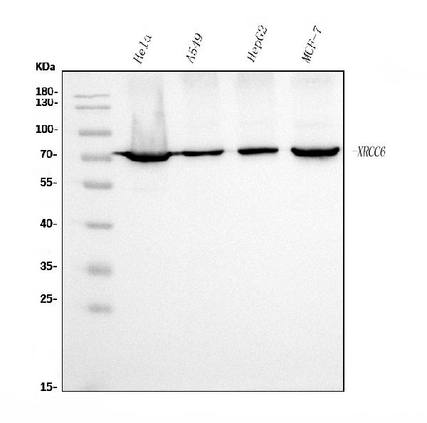
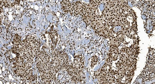
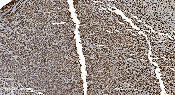
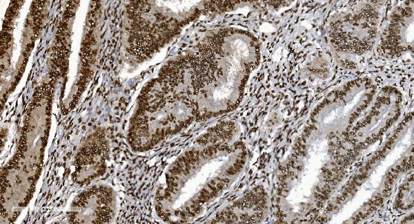
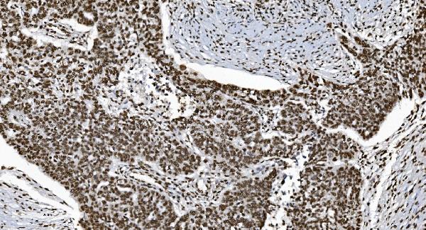
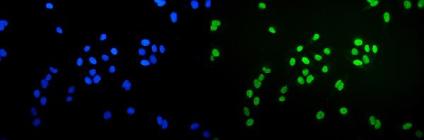
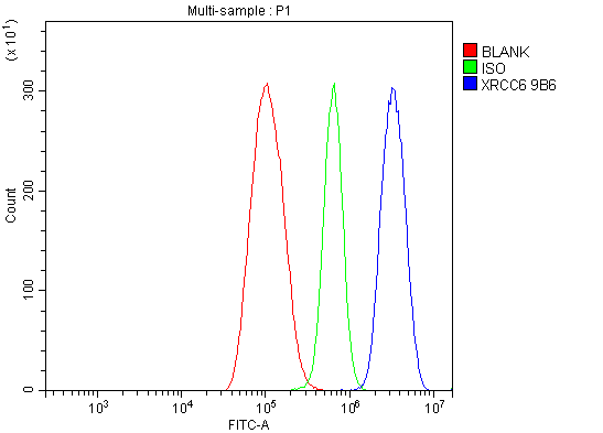
Anti-Ku70 Antibody Picoband™ (monoclonal, 9B6)
- SPECIFICATION
- CITATIONS
- PROTOCOLS
- BACKGROUND

Application
| WB, IHC, IF, ICC, FC |
|---|---|
| Primary Accession | P12956 |
| Host | Mouse |
| Isotype | Mouse IgG2b |
| Reactivity | Human |
| Clonality | Monoclonal |
| Format | Lyophilized |
| Description | Anti-Ku70 Antibody Picoband™ (monoclonal, 9B6) . Tested in Flow Cytometry, IF, IHC, ICC, WB applications. This antibody reacts with Human. |
| Reconstitution | Add 0.2ml of distilled water will yield a concentration of 500ug/ml. |
| Gene ID | 2547 |
|---|---|
| Other Names | X-ray repair cross-complementing protein 6, 3.6.4.-, 4.2.99.-, 5'-deoxyribose-5-phosphate lyase Ku70, 5'-dRP lyase Ku70, 70 kDa subunit of Ku antigen, ATP-dependent DNA helicase 2 subunit 1, ATP-dependent DNA helicase II 70 kDa subunit, CTC box-binding factor 75 kDa subunit, CTC75, CTCBF, DNA repair protein XRCC6, Lupus Ku autoantigen protein p70, Ku70, Thyroid-lupus autoantigen, TLAA, X-ray repair complementing defective repair in Chinese hamster cells 6, XRCC6, G22P1 |
| Calculated MW | 70 kDa |
| Application Details | Western blot, 0.25-0.5 µg/ml, Human Immunohistochemistry (Paraffin-embedded Section), 2-5 µg/ml, Human Immunocytochemistry/Immunofluorescence, 5 µg/ml, Human Flow Cytometry, 1-3 µg/1x10^6 cells, Human |
| Contents | Each vial contains 4mg Trehalose, 0.9mg NaCl and 0.2mg Na2HPO4. |
| Clone Names | Clone: 9B6 |
| Immunogen | A synthetic peptide corresponding to a sequence at C-terminus of human Ku70, different from the related mouse sequence by one amino acid. |
| Purification | Immunogen affinity purified. |
| Storage | Store at -20˚C for one year from date of receipt. After reconstitution, at 4˚C for one month. It can also be aliquotted and stored frozen at -20˚C for six months. Avoid repeated freeze-thaw cycles. |
| Name | XRCC6 |
|---|---|
| Synonyms | G22P1 |
| Function | Single-stranded DNA-dependent ATP-dependent helicase that plays a key role in DNA non-homologous end joining (NHEJ) by recruiting DNA-PK to DNA (PubMed:11493912, PubMed:12145306, PubMed:20493174, PubMed:2466842, PubMed:7957065, PubMed:8621488, PubMed:9742108). Required for double-strand break repair and V(D)J recombination (PubMed:11493912, PubMed:12145306, PubMed:20493174, PubMed:2466842, PubMed:7957065, PubMed:8621488, PubMed:9742108). Also has a role in chromosome translocation (PubMed:11493912, PubMed:12145306, PubMed:20493174, PubMed:2466842, PubMed:7957065, PubMed:8621488, PubMed:9742108). Has a role in chromosome translocation (PubMed:11493912, PubMed:12145306, PubMed:20493174, PubMed:2466842, PubMed:7957065, PubMed:8621488, PubMed:9742108). The DNA helicase II complex binds preferentially to fork-like ends of double-stranded DNA in a cell cycle-dependent manner (PubMed:11493912, PubMed:12145306, PubMed:20493174, PubMed:2466842, PubMed:7957065, PubMed:8621488, PubMed:9742108). It works in the 3'-5' direction (PubMed:11493912, PubMed:12145306, PubMed:20493174, PubMed:2466842, PubMed:7957065, PubMed:8621488, PubMed:9742108). During NHEJ, the XRCC5-XRRC6 dimer performs the recognition step: it recognizes and binds to the broken ends of the DNA and protects them from further resection (PubMed:11493912, PubMed:12145306, PubMed:20493174, PubMed:2466842, PubMed:7957065, PubMed:8621488, PubMed:9742108). Binding to DNA may be mediated by XRCC6 (PubMed:11493912, PubMed:12145306, PubMed:20493174, PubMed:2466842, PubMed:7957065, PubMed:8621488, PubMed:9742108). The XRCC5-XRRC6 dimer acts as a regulatory subunit of the DNA-dependent protein kinase complex DNA-PK by increasing the affinity of the catalytic subunit PRKDC to DNA by 100-fold (PubMed:11493912, PubMed:12145306, PubMed:20493174, PubMed:2466842, PubMed:7957065, PubMed:8621488, PubMed:9742108). The XRCC5-XRRC6 dimer is probably involved in stabilizing broken DNA ends and bringing them together (PubMed:11493912, PubMed:12145306, PubMed:20493174, PubMed:2466842, PubMed:7957065, PubMed:8621488, PubMed:9742108). The assembly of the DNA-PK complex to DNA ends is required for the NHEJ ligation step (PubMed:11493912, PubMed:12145306, PubMed:20493174, PubMed:2466842, PubMed:7957065, PubMed:8621488, PubMed:9742108). Probably also acts as a 5'-deoxyribose-5-phosphate lyase (5'-dRP lyase), by catalyzing the beta-elimination of the 5' deoxyribose-5-phosphate at an abasic site near double-strand breaks (PubMed:20383123). 5'-dRP lyase activity allows to 'clean' the termini of abasic sites, a class of nucleotide damage commonly associated with strand breaks, before such broken ends can be joined (PubMed:20383123). The XRCC5-XRRC6 dimer together with APEX1 acts as a negative regulator of transcription (PubMed:8621488). In association with NAA15, the XRCC5-XRRC6 dimer binds to the osteocalcin promoter and activates osteocalcin expression (PubMed:12145306). Plays a role in the regulation of DNA virus-mediated innate immune response by assembling into the HDP-RNP complex, a complex that serves as a platform for IRF3 phosphorylation and subsequent innate immune response activation through the cGAS-STING pathway (PubMed:28712728). |
| Cellular Location | Nucleus. Chromosome |

Thousands of laboratories across the world have published research that depended on the performance of antibodies from Abcepta to advance their research. Check out links to articles that cite our products in major peer-reviewed journals, organized by research category.
info@abcepta.com, and receive a free "I Love Antibodies" mug.

Provided below are standard protocols that you may find useful for product applications.
Background
XRCC6 (X-Ray Repair, Complementing Defective, In Chinese Hamster, 6), also called Ku70, G22P1 or TLAA, is a protein that in humans, is encoded by the XRCC6 gene. In addition, the XRCC6 gene encodes subunit p70 of the p70/p80 autoantigen which consists of 2 proteins of molecular mass of approximately 70,000 and 80,000 daltons that dimerize to form a 10 S DNA-binding complex. The XRCC6 gene is mapped to 22q13.2. XRCC6 and Mre11 are differentially expressed during meiosis. XRCC6 interacts with Baxa, a mediator of mitochondrial-dependent apoptosis. Disruption of both FANCC and XRCC6 suppressed sensitivity to crosslinking agents, diminished chromosome breaks, and reversed defective homologous recombination. Ku70 binds directly to free DNA ends, committing them to NHEJ repair. In early meiotic prophase, however, when meiotic recombination is most probably initiated, Mre11 was abundant, whereas XRCC6 was not detectable.
If you have used an Abcepta product and would like to share how it has performed, please click on the "Submit Review" button and provide the requested information. Our staff will examine and post your review and contact you if needed.
If you have any additional inquiries please email technical services at tech@abcepta.com.
Ordering Information
Other Products
Shipping Information
