


 Foundational characteristics of cancer include proliferation, angiogenesis, migration, evasion of apoptosis, and cellular immortality. Find key markers for these cellular processes and antibodies to detect them.
 The SUMOplot™ Analysis Program predicts and scores sumoylation sites in your protein. SUMOylation is a post-translational modification involved in various cellular processes, such as nuclear-cytosolic transport, transcriptional regulation, apoptosis, protein stability, response to stress, and progression through the cell cycle.
 The Autophagy Receptor Motif Plotter predicts and scores autophagy receptor binding sites in your protein. Identifying proteins connected to this pathway is critical to understanding the role of autophagy in physiological as well as pathological processes such as development, differentiation, neurodegenerative diseases, stress, infection, and cancer.
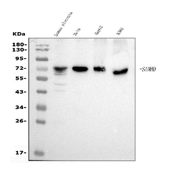
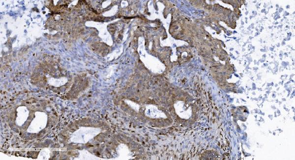
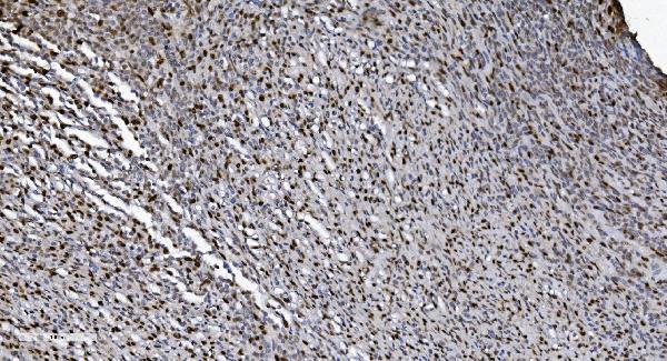
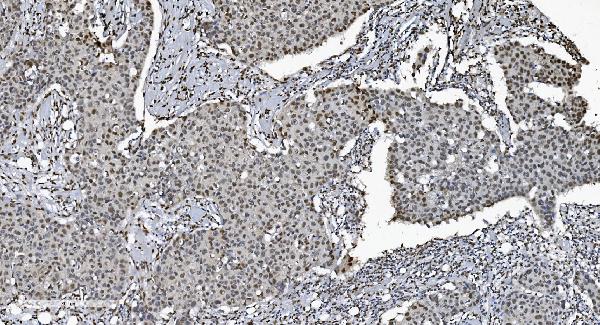
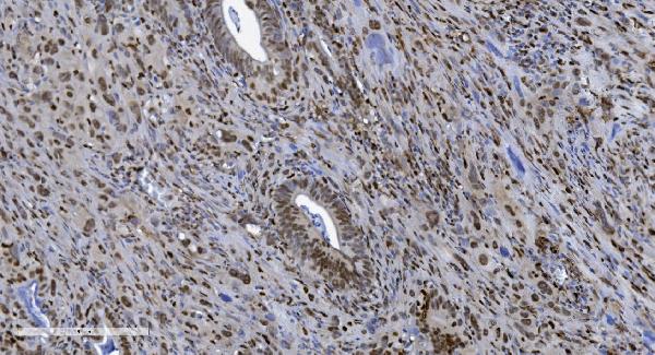
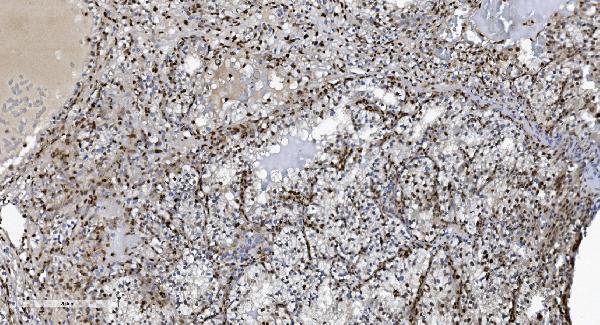
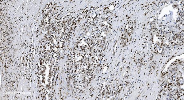
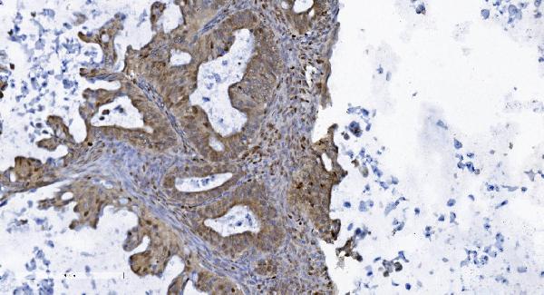
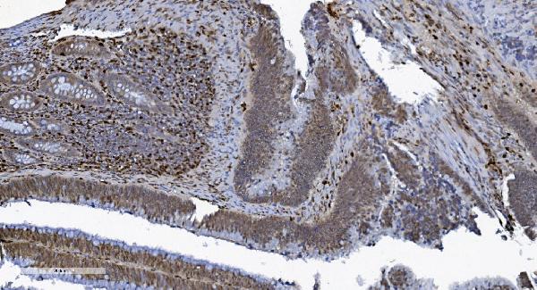
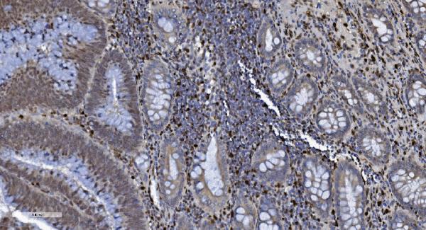
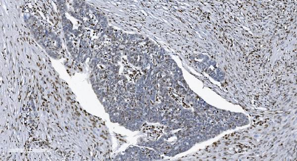
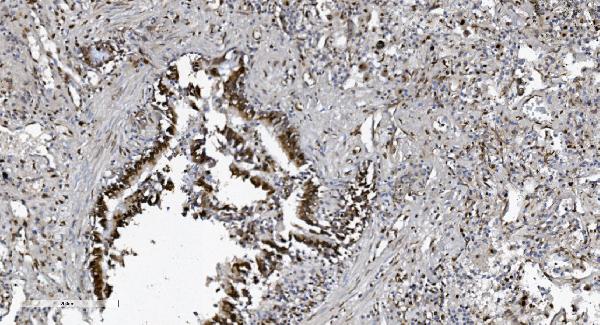
Anti-SAMHD1 Antibody Picoband™ (monoclonal, 10H8)
- SPECIFICATION
- CITATIONS
- PROTOCOLS
- BACKGROUND

Application
| WB, IHC, IF, ICC, FC |
|---|---|
| Primary Accession | Q9Y3Z3 |
| Host | Mouse |
| Isotype | Mouse IgG2b |
| Reactivity | Human |
| Clonality | Monoclonal |
| Format | Lyophilized |
| Description | Anti-SAMHD1 Antibody Picoband™ (monoclonal, 10H8) . Tested in Flow Cytometry, IF, IHC, ICC, WB applications. This antibody reacts with Human. |
| Reconstitution | Add 0.2ml of distilled water will yield a concentration of 500ug/ml. |
| Gene ID | 25939 |
|---|---|
| Other Names | Deoxynucleoside triphosphate triphosphohydrolase SAMHD1, dNTPase, 3.1.5.-, Dendritic cell-derived IFNG-induced protein, DCIP, Monocyte protein 5 {ECO:0000303|Ref.2}, MOP-5 {ECO:0000303|Ref.2}, SAM domain and HD domain-containing protein 1, hSAMHD1, SAMHD1 (HGNC:15925) |
| Calculated MW | 72 kDa |
| Application Details | Western blot, 0.25-0.5 µg/ml, Human Immunohistochemistry (Paraffin-embedded Section), 2-5 µg/ml, Human Immunocytochemistry/Immunofluorescence, 5 µg/ml, Human Flow Cytometry, 1-3 µg/1x10^6 cells, Human |
| Contents | Each vial contains 4mg Trehalose, 0.9mg NaCl and 0.2mg Na2HPO4. |
| Clone Names | Clone: 10H8 |
| Immunogen | E.coli-derived human SAMHD1 recombinant protein (Position: E37-M626). |
| Purification | Immunogen affinity purified. |
| Storage | Store at -20˚C for one year from date of receipt. After reconstitution, at 4˚C for one month. It can also be aliquotted and stored frozen at -20˚C for six months. Avoid repeated freeze-thaw cycles. |
| Name | SAMHD1 (HGNC:15925) |
|---|---|
| Function | Protein that acts both as a host restriction factor involved in defense response to virus and as a regulator of DNA end resection at stalled replication forks (PubMed:19525956, PubMed:21613998, PubMed:21720370, PubMed:22056990, PubMed:23601106, PubMed:23602554, PubMed:24336198, PubMed:26294762, PubMed:26431200, PubMed:28229507, PubMed:28834754, PubMed:29670289). Has deoxynucleoside triphosphate (dNTPase) activity, which is required to restrict infection by viruses, such as HIV-1: dNTPase activity reduces cellular dNTP levels to levels too low for retroviral reverse transcription to occur, blocking early- stage virus replication in dendritic and other myeloid cells (PubMed:19525956, PubMed:21613998, PubMed:21720370, PubMed:22056990, PubMed:23364794, PubMed:23601106, PubMed:23602554, PubMed:24336198, PubMed:25038827, PubMed:26101257, PubMed:26294762, PubMed:26431200, PubMed:28229507). Likewise, suppresses LINE-1 retrotransposon activity (PubMed:24035396, PubMed:24217394, PubMed:29610582). Not able to restrict infection by HIV-2 virus; because restriction activity is counteracted by HIV-2 viral protein Vpx (PubMed:21613998, PubMed:21720370). In addition to virus restriction, dNTPase activity acts as a regulator of DNA precursor pools by regulating dNTP pools (PubMed:23858451). Phosphorylation at Thr-592 acts as a switch to control dNTPase-dependent and -independent functions: it inhibits dNTPase activity and ability to restrict infection by viruses, while it promotes DNA end resection at stalled replication forks (PubMed:23601106, PubMed:23602554, PubMed:29610582, PubMed:29670289). Functions during S phase at stalled DNA replication forks to promote the resection of gapped or reversed forks: acts by stimulating the exonuclease activity of MRE11, activating the ATR-CHK1 pathway and allowing the forks to restart replication (PubMed:29670289). Its ability to promote degradation of nascent DNA at stalled replication forks is required to prevent induction of type I interferons, thereby preventing chronic inflammation (PubMed:27477283, PubMed:29670289). Ability to promote DNA end resection at stalled replication forks is independent of dNTPase activity (PubMed:29670289). Enhances immunoglobulin hypermutation in B-lymphocytes by promoting transversion mutation (By similarity). |
| Cellular Location | Nucleus. Chromosome Note=Localizes to sites of DNA double-strand breaks in response to DNA damage. |
| Tissue Location | Expressed in heart, skeletal muscle, spleen, liver, small intestine, placenta, lung and peripheral blood leukocytes (PubMed:11064105). No expression is seen in brain and thymus (PubMed:11064105). |

Thousands of laboratories across the world have published research that depended on the performance of antibodies from Abcepta to advance their research. Check out links to articles that cite our products in major peer-reviewed journals, organized by research category.
info@abcepta.com, and receive a free "I Love Antibodies" mug.

Provided below are standard protocols that you may find useful for product applications.
Background
SAM domain and HD domain-containing protein 1 is a protein that in humans is encoded by the SAMHD1 gene. This gene may play a role in regulation of the innate immune response. The encoded protein is upregulated in response to viral infection and may be involved in mediation of tumor necrosis factor-alpha proinflammatory responses. Mutations in this gene have been associated with Aicardi-Goutieres syndrome.
If you have used an Abcepta product and would like to share how it has performed, please click on the "Submit Review" button and provide the requested information. Our staff will examine and post your review and contact you if needed.
If you have any additional inquiries please email technical services at tech@abcepta.com.
Ordering Information
Other Products
Shipping Information
