


 Foundational characteristics of cancer include proliferation, angiogenesis, migration, evasion of apoptosis, and cellular immortality. Find key markers for these cellular processes and antibodies to detect them.
 The SUMOplot™ Analysis Program predicts and scores sumoylation sites in your protein. SUMOylation is a post-translational modification involved in various cellular processes, such as nuclear-cytosolic transport, transcriptional regulation, apoptosis, protein stability, response to stress, and progression through the cell cycle.
 The Autophagy Receptor Motif Plotter predicts and scores autophagy receptor binding sites in your protein. Identifying proteins connected to this pathway is critical to understanding the role of autophagy in physiological as well as pathological processes such as development, differentiation, neurodegenerative diseases, stress, infection, and cancer.
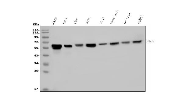
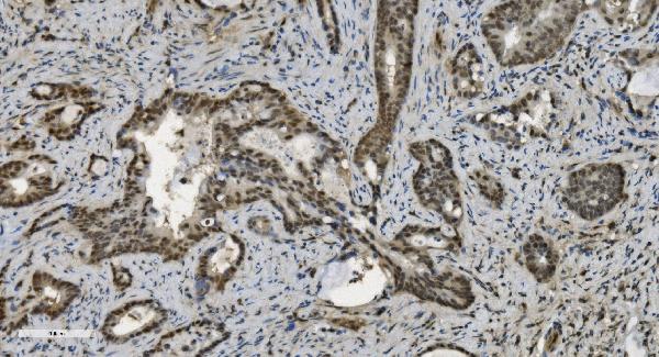
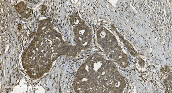
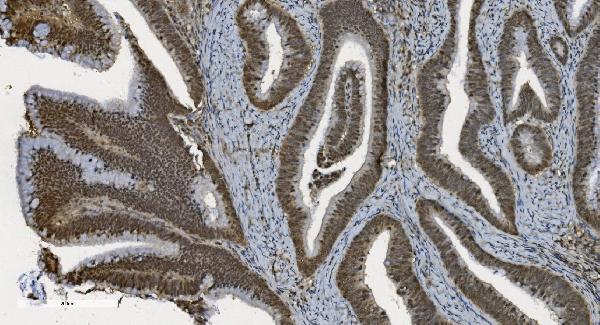
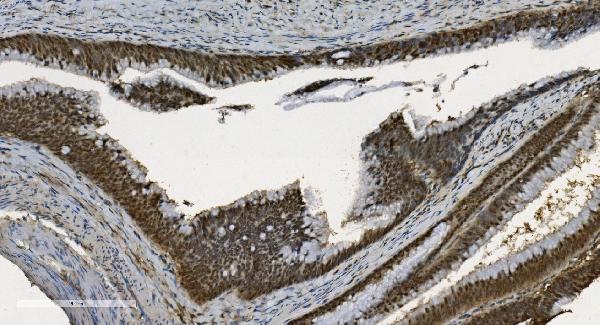
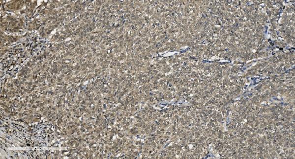
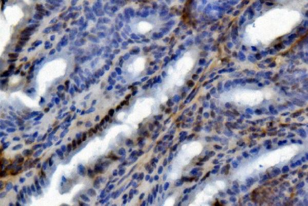
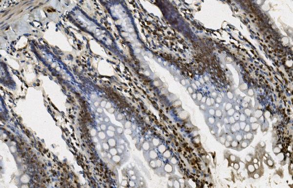
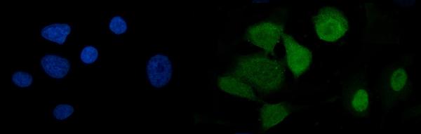
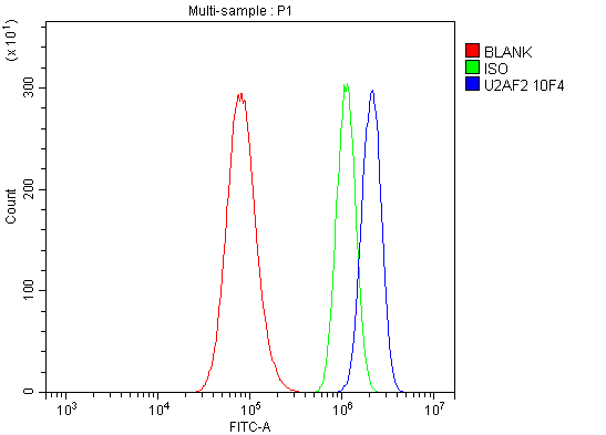
Anti-U2AF65/U2AF2 Picoband™ Antibody (monoclonal, 10F4)
- SPECIFICATION
- CITATIONS
- PROTOCOLS
- BACKGROUND

Application
| WB, IHC, IF, ICC, FC |
|---|---|
| Primary Accession | P26368 |
| Host | Mouse |
| Isotype | Mouse IgG2b |
| Reactivity | Rat, Human, Mouse |
| Clonality | Monoclonal |
| Format | Lyophilized |
| Description | Anti-U2AF65/U2AF2 Picoband™ Antibody (monoclonal, 10F4) . Tested in Flow Cytometry, IF, IHC, ICC, WB applications. This antibody reacts with Human, Mouse, Rat. |
| Reconstitution | Add 0.2ml of distilled water will yield a concentration of 500ug/ml. |
| Gene ID | 11338 |
|---|---|
| Other Names | Splicing factor U2AF 65 kDa subunit, U2 auxiliary factor 65 kDa subunit, hU2AF(65), hU2AF65, U2 snRNP auxiliary factor large subunit, U2AF2, U2AF65 |
| Calculated MW | 65 kDa |
| Application Details | Western blot, 0.1-0.25 µg/ml, Human, Mouse, Rat Immunohistochemistry (Paraffin-embedded Section), 2-5 µg/ml, Human, Mouse, Rat Immunocytochemistry/Immunofluorescence, 5 µg/ml, Human Flow Cytometry, 1-3 µg/1x10^6 cells, Human |
| Contents | Each vial contains 4mg Trehalose, 0.9mg NaCl and 0.2mg Na2HPO4. |
| Clone Names | Clone: 10F4 |
| Immunogen | E.coli-derived human U2AF65/U2AF2 recombinant protein (Position: M238-H470). |
| Purification | Immunogen affinity purified. |
| Storage | Store at -20˚C for one year from date of receipt. After reconstitution, at 4˚C for one month. It can also be aliquotted and stored frozen at -20˚C for six months. Avoid repeated freeze-thaw cycles. |
| Name | U2AF2 |
|---|---|
| Synonyms | U2AF65 |
| Function | Plays a role in pre-mRNA splicing and 3'-end processing (PubMed:17024186). By recruiting PRPF19 and the PRP19C/Prp19 complex/NTC/Nineteen complex to the RNA polymerase II C-terminal domain (CTD), and thereby pre-mRNA, may couple transcription to splicing (PubMed:21536736). Induces cardiac troponin-T (TNNT2) pre-mRNA exon inclusion in muscle. Regulates the TNNT2 exon 5 inclusion through competition with MBNL1. Binds preferentially to a single-stranded structure within the polypyrimidine tract of TNNT2 intron 4 during spliceosome assembly. Required for the export of mRNA out of the nucleus, even if the mRNA is encoded by an intron-less gene. Represses the splicing of MAPT/Tau exon 10. Positively regulates pre-mRNA 3'-end processing by recruiting the CFIm complex to cleavage and polyadenylation signals (PubMed:17024186). |
| Cellular Location | Nucleus. |

Thousands of laboratories across the world have published research that depended on the performance of antibodies from Abcepta to advance their research. Check out links to articles that cite our products in major peer-reviewed journals, organized by research category.
info@abcepta.com, and receive a free "I Love Antibodies" mug.

Provided below are standard protocols that you may find useful for product applications.
Background
Splicing factor U2AF 65 kDa subunit is a protein that in humans is encoded by the U2AF2 gene. It is mapped to 19q13.42. U2 auxiliary factor (U2AF), comprised of a large and a small subunit, is a non-snRNP protein required for the binding of U2 snRNP to the pre-mRNA branch site. This gene encodes the U2AF large subunit which contains a sequence-specific RNA-binding region with 3 RNA recognition motifs and an Arg/Ser-rich domain necessary for splicing. The large subunit binds to the polypyrimidine tract of introns early during spliceosome assembly. Multiple transcript variants have been detected for this gene, but the full-length natures of only two have been determined to date.
If you have used an Abcepta product and would like to share how it has performed, please click on the "Submit Review" button and provide the requested information. Our staff will examine and post your review and contact you if needed.
If you have any additional inquiries please email technical services at tech@abcepta.com.
Ordering Information
Other Products
Shipping Information
