


 Foundational characteristics of cancer include proliferation, angiogenesis, migration, evasion of apoptosis, and cellular immortality. Find key markers for these cellular processes and antibodies to detect them.
 The SUMOplot™ Analysis Program predicts and scores sumoylation sites in your protein. SUMOylation is a post-translational modification involved in various cellular processes, such as nuclear-cytosolic transport, transcriptional regulation, apoptosis, protein stability, response to stress, and progression through the cell cycle.
 The Autophagy Receptor Motif Plotter predicts and scores autophagy receptor binding sites in your protein. Identifying proteins connected to this pathway is critical to understanding the role of autophagy in physiological as well as pathological processes such as development, differentiation, neurodegenerative diseases, stress, infection, and cancer.
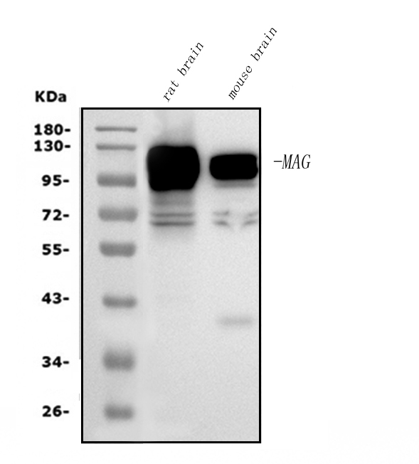
Anti-MAG Picoband™ Antibody (monoclonal, 2G11)
- SPECIFICATION
- CITATIONS
- PROTOCOLS
- BACKGROUND

Application
| WB, IHC, IF, FC |
|---|---|
| Primary Accession | P20916 |
| Host | Mouse |
| Isotype | Mouse IgG2a |
| Reactivity | Rat, Human, Mouse |
| Clonality | Monoclonal |
| Format | Lyophilized |
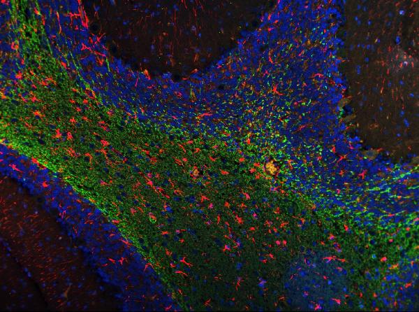
| Description | Anti-MAG Picoband™ Antibody (monoclonal, 2G11) . Tested in Flow Cytometry, IF, IHC, WB applications. This antibody reacts with Human, Mouse, Rat. |
| Reconstitution | Add 0.2ml of distilled water will yield a concentration of 500ug/ml. |
| Gene ID | 4099 |
|---|---|
| Other Names | Myelin-associated glycoprotein, Siglec-4a, MAG, GMA |
| Calculated MW | 100 kDa |
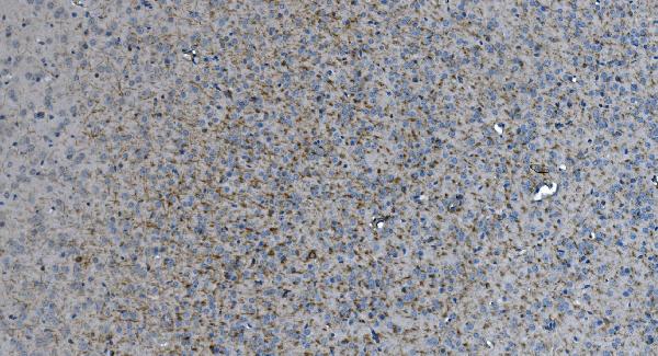
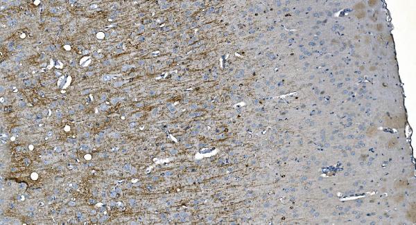
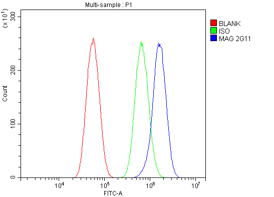
| Application Details | Western blot, 0.1-0.25 µg/ml, Mouse, Rat Immunohistochemistry (Paraffin-embedded Section), 2-5 µg/ml, Mouse,Rat Immunofluorescence, 5 µg/ml, Rat Flow Cytometry, 1-3 µg/1x10^6 cells, Human |
| Contents | Each vial contains 4mg Trehalose, 0.9mg NaCl and 0.2mg Na2HPO4. |
| Clone Names | Clone: 2G11 |
| Immunogen | E.coli-derived human MAG recombinant protein (Position: E34-R605). |
| Purification | Immunogen affinity purified. |
| Storage | Store at -20˚C for one year from date of receipt. After reconstitution, at 4˚C for one month. It can also be aliquotted and stored frozen at -20˚C for six months. Avoid repeated freeze-thaw cycles. |
| Name | MAG |
|---|---|
| Synonyms | GMA |
| Function | Adhesion molecule that mediates interactions between myelinating cells and neurons by binding to neuronal sialic acid- containing gangliosides and to the glycoproteins RTN4R and RTN4RL2 (By similarity). Not required for initial myelination, but seems to play a role in the maintenance of normal axon myelination. Protects motoneurons against apoptosis, also after injury; protection against apoptosis is probably mediated via interaction with neuronal RTN4R and RTN4RL2. Required to prevent degeneration of myelinated axons in adults; this probably depends on binding to gangliosides on the axon cell membrane (By similarity). Negative regulator of neurite outgrowth; in dorsal root ganglion neurons the inhibition is mediated primarily via binding to neuronal RTN4R or RTN4RL2 and to a lesser degree via binding to neuronal gangliosides. In cerebellar granule cells the inhibition is mediated primarily via binding to neuronal gangliosides. In sensory neurons, inhibition of neurite extension depends only partially on RTN4R, RTN4RL2 and gangliosides. Inhibits axon longitudinal growth (By similarity). Inhibits axon outgrowth by binding to RTN4R (By similarity). Preferentially binds to alpha-2,3-linked sialic acid. Binds ganglioside Gt1b (By similarity). |
| Cellular Location | Cell membrane; Single-pass type I membrane protein Membrane raft {ECO:0000250|UniProtKB:P07722} |
| Tissue Location | Both isoform 1 and isoform 2 are detected in myelinated structures in the central and peripheral nervous system, in periaxonal myelin and at Schmidt-Lanterman incisures (PubMed:6200494, PubMed:9495552). Detected in optic nerve, in oligodendroglia and in periaxonal myelin sheaths (PubMed:6200494). Detected in compact myelin (at protein level) (PubMed:6200494). Both isoform 1 and isoform 2 are detected in the central and peripheral nervous system (PubMed:9495552) |

Thousands of laboratories across the world have published research that depended on the performance of antibodies from Abcepta to advance their research. Check out links to articles that cite our products in major peer-reviewed journals, organized by research category.
info@abcepta.com, and receive a free "I Love Antibodies" mug.

Provided below are standard protocols that you may find useful for product applications.
Background
MAG (Myelin-associated glycoprotein),also known as SIGLEC4A, is a cell membrane glycoprotein that is a member of the SIGLEC family of proteins and is a functional ligand of the NOGO-66 receptor, NgR. It is though to be involved in the process of myelination. MAG is a sialic acid-binding SIGLEC protein and is a functional ligand for the NOGO receptor.The MAG gene is mapped on 19q13.12. Cleavage of GPI-linked proteins from axons protects growth cones from MAG-induced collapse, and dominant-negative NgR eliminates MAG inhibition of neurite outgrowth. MAG-resistant embryonic neurons were rendered MAG-sensitive by expression of NgR. MAG binds specifically to an NgR-expressing cell line in a GPI-dependent and sialic acid-independent manner. Experiments blocking NgR from interacting with MAG prevented inhibition of neurite outgrowth by MAG. In cultured human embryonic kidney (HEK) cells expressing the NOGO receptor, p75 (NTR) was required for MAG-induced intracellular calcium elevation.
If you have used an Abcepta product and would like to share how it has performed, please click on the "Submit Review" button and provide the requested information. Our staff will examine and post your review and contact you if needed.
If you have any additional inquiries please email technical services at tech@abcepta.com.
Ordering Information
Other Products
Shipping Information
