


 Foundational characteristics of cancer include proliferation, angiogenesis, migration, evasion of apoptosis, and cellular immortality. Find key markers for these cellular processes and antibodies to detect them.
 The SUMOplot™ Analysis Program predicts and scores sumoylation sites in your protein. SUMOylation is a post-translational modification involved in various cellular processes, such as nuclear-cytosolic transport, transcriptional regulation, apoptosis, protein stability, response to stress, and progression through the cell cycle.
 The Autophagy Receptor Motif Plotter predicts and scores autophagy receptor binding sites in your protein. Identifying proteins connected to this pathway is critical to understanding the role of autophagy in physiological as well as pathological processes such as development, differentiation, neurodegenerative diseases, stress, infection, and cancer.
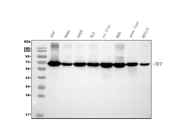
Anti-Transketolase/TKT Picoband™ Antibody (monoclonal, 3E5)
- SPECIFICATION
- CITATIONS
- PROTOCOLS
- BACKGROUND

Application
| WB, IHC, IF, ICC, FC |
|---|---|
| Primary Accession | P29401 |
| Host | Mouse |
| Isotype | Mouse IgG2a |
| Reactivity | Rat, Human, Mouse |
| Clonality | Monoclonal |
| Format | Lyophilized |
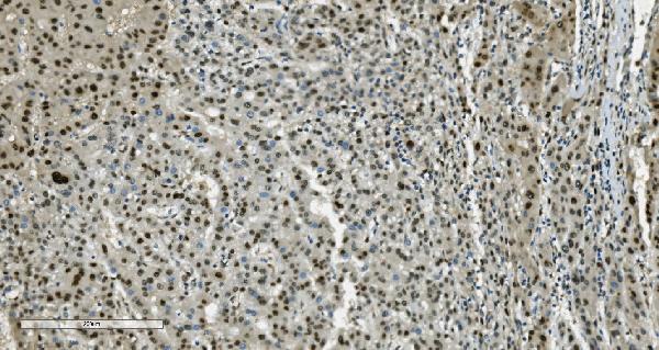
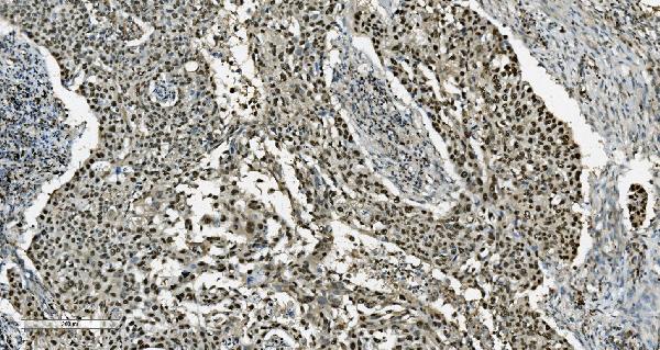
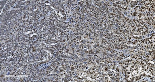
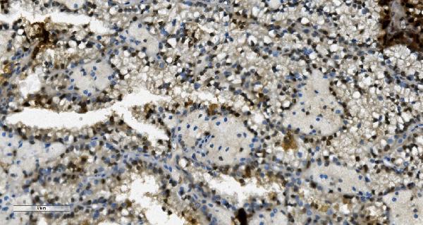
| Description | Anti-Transketolase/TKT Picoband™ Antibody (monoclonal, 3E5) . Tested in Flow Cytometry, IF, IHC, ICC, WB applications. This antibody reacts with Human, Mouse, Rat. |
| Reconstitution | Add 0.2ml of distilled water will yield a concentration of 500ug/ml. |
| Gene ID | 7086 |
|---|---|
| Other Names | Transketolase, TK, 2.2.1.1, TKT |
| Calculated MW | 68 kDa |
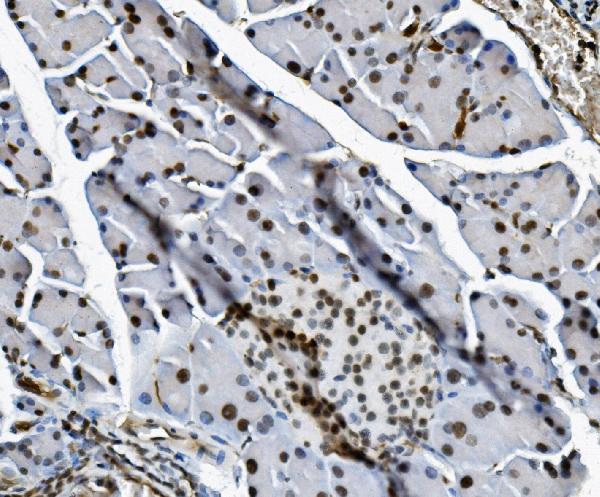
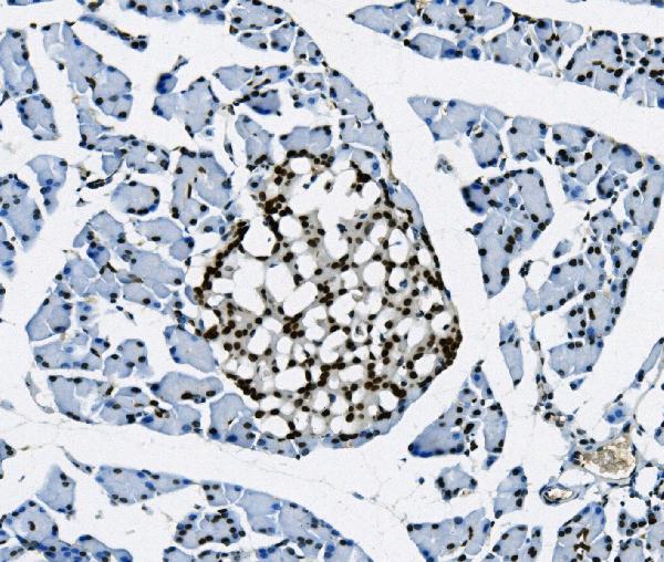
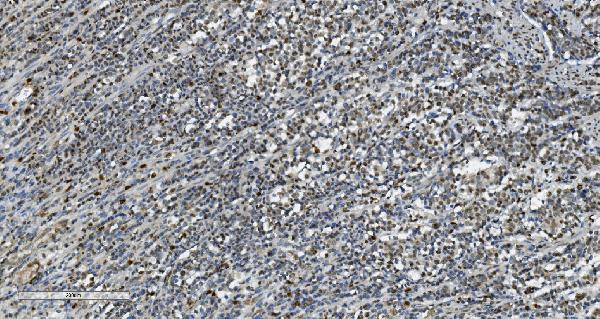
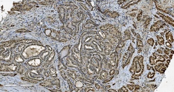
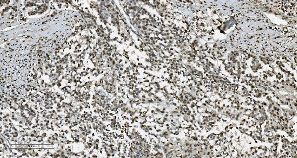
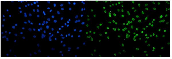
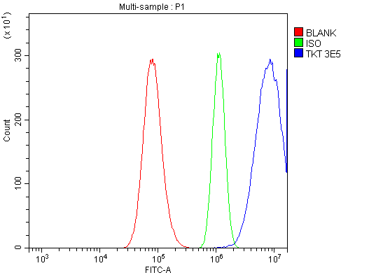
| Application Details | Western blot, 0.1-0.25 µg/ml, Human, Mouse, Rat Immunohistochemistry (Paraffin-embedded Section), 1-2 µg/ml, Human, Mouse, Rat Immunocytochemistry/Immunofluorescence; 5 µg/ml, Human Flow Cytometry, 1-3 µg/1x10^6 cells, Human |
| Contents | Each vial contains 4mg Trehalose, 0.9mg NaCl and 0.2mg Na2HPO4. |
| Clone Names | Clone: 3E5 |
| Immunogen | E.coli-derived human Transketolase/TKT recombinant protein (Position: M1-A116). |
| Purification | Immunogen affinity purified. |
| Storage | Store at -20˚C for one year from date of receipt. After reconstitution, at 4˚C for one month. It can also be aliquotted and stored frozen at -20˚C for six months. Avoid repeated freeze-thaw cycles. |
| Name | TKT |
|---|---|
| Function | Catalyzes the transfer of a two-carbon ketol group from a ketose donor to an aldose acceptor, via a covalent intermediate with the cofactor thiamine pyrophosphate. |

Thousands of laboratories across the world have published research that depended on the performance of antibodies from Abcepta to advance their research. Check out links to articles that cite our products in major peer-reviewed journals, organized by research category.
info@abcepta.com, and receive a free "I Love Antibodies" mug.

Provided below are standard protocols that you may find useful for product applications.
Background
Transketolase is a thiamine-dependent enzyme that links the pentose phosphate pathway with the glycolytic pathway. The pentose phosphate pathway, which is active in most tissues, provides sugar phosphates for intermediary biosynthesis, especially nucleotide metabolism, and generates the biosynthetic reducing power for the cell in the form of NADPH. Transketolase is directly involved in the branch of the pathway that channels excess sugar phosphates to glycolysis, enabling the production of NADPH to be maintained under different metabolic conditions. NADPH is critical for maintaining cerebral glutathione, and thus it is likely that transketolase plays an important role in brain metabolism.
If you have used an Abcepta product and would like to share how it has performed, please click on the "Submit Review" button and provide the requested information. Our staff will examine and post your review and contact you if needed.
If you have any additional inquiries please email technical services at tech@abcepta.com.
Ordering Information
Other Products
Shipping Information
