


 Foundational characteristics of cancer include proliferation, angiogenesis, migration, evasion of apoptosis, and cellular immortality. Find key markers for these cellular processes and antibodies to detect them.
 The SUMOplot™ Analysis Program predicts and scores sumoylation sites in your protein. SUMOylation is a post-translational modification involved in various cellular processes, such as nuclear-cytosolic transport, transcriptional regulation, apoptosis, protein stability, response to stress, and progression through the cell cycle.
 The Autophagy Receptor Motif Plotter predicts and scores autophagy receptor binding sites in your protein. Identifying proteins connected to this pathway is critical to understanding the role of autophagy in physiological as well as pathological processes such as development, differentiation, neurodegenerative diseases, stress, infection, and cancer.
Anti-CDC45L Picoband™ Antibody (monoclonal, 6H6)


- SPECIFICATION
- CITATIONS
- PROTOCOLS
- BACKGROUND

Application
| WB, IHC, FC |
|---|---|
| Primary Accession | O75419 |
| Host | Mouse |
| Isotype | Mouse IgG1 |
| Reactivity | Rat, Human, Mouse |
| Clonality | Monoclonal |
| Format | Lyophilized |
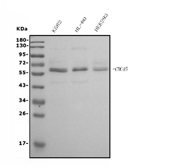
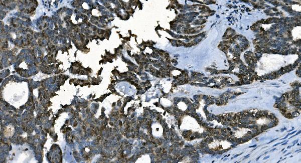
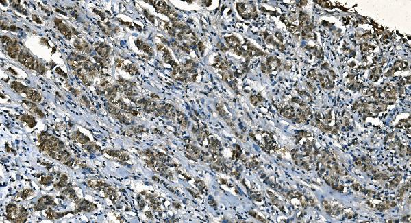
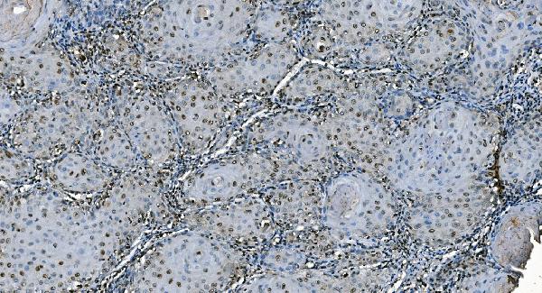
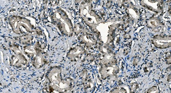
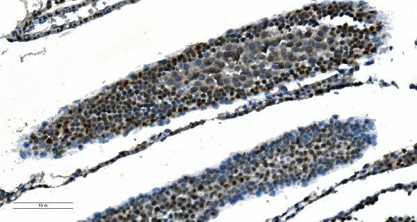
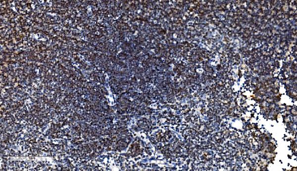
| Description | Anti-CDC45L Picoband™ Antibody (monoclonal, 6H6) . Tested in Flow Cytometry, IHC, WB applications. This antibody reacts with Human, Mouse, Rat. |
| Reconstitution | Add 0.2ml of distilled water will yield a concentration of 500ug/ml. |
| Gene ID | 8318 |
|---|---|
| Other Names | Cell division control protein 45 homolog, PORC-PI-1, CDC45 (HGNC:1739), CDC45L, CDC45L2 |
| Calculated MW | 66 kDa |
| Application Details | Western blot, 0.25-0.5 µg/ml, Human Immunohistochemistry (Paraffin-embedded Section), 2-5 µg/ml, Human, Mouse, Rat Flow Cytometry, 1-3 µg/1x10^6 cells, Human |
| Contents | Each vial contains 4mg Trehalose, 0.9mg NaCl and 0.2mg Na2HPO4. |
| Clone Names | Clone: 6H6 |
| Immunogen | E. coli-derived human CDC45L recombinant protein (Position: E166-A431). |
| Purification | Immunogen affinity purified. |
| Storage | Store at -20˚C for one year from date of receipt. After reconstitution, at 4˚C for one month. It can also be aliquotted and stored frozen at -20˚C for six months. Avoid repeated freeze-thaw cycles. |
| Name | CDC45 (HGNC:1739) |
|---|---|
| Synonyms | CDC45L, CDC45L2 |
| Function | Required for initiation of chromosomal DNA replication. Core component of CDC45-MCM-GINS (CMG) helicase, the molecular machine that unwinds template DNA during replication, and around which the replisome is built. |
| Cellular Location | Nucleus. Chromosome. Note=Associates with chromatin |
| Tissue Location | Widely expressed, highest levels are found in adult testis and thymus and in fetal liver |

Thousands of laboratories across the world have published research that depended on the performance of antibodies from Abcepta to advance their research. Check out links to articles that cite our products in major peer-reviewed journals, organized by research category.
info@abcepta.com, and receive a free "I Love Antibodies" mug.

Provided below are standard protocols that you may find useful for product applications.
Background
CDC45 is a protein that in humans is encoded by the CDC45L gene. The protein encoded by this gene was identified by its strong similarity with Saccharomyces cerevisiae Cdc45, an essential protein required to the initiation of DNA replication. Cdc45 is a member of the highly conserved multiprotein complex including Cdc6/Cdc18, the minichromosome maintenance proteins (MCMs) and DNA polymerase, which is important for early steps of DNA replication in eukaryotes. This protein has been shown to interact with MCM7 and DNA polymerase alpha. Studies of the similar gene in Xenopus suggested that this protein play a pivotal role in the loading of DNA polymerase alpha onto chromatin. Alternate splicing results in multiple transcript variants.
If you have used an Abcepta product and would like to share how it has performed, please click on the "Submit Review" button and provide the requested information. Our staff will examine and post your review and contact you if needed.
If you have any additional inquiries please email technical services at tech@abcepta.com.
Ordering Information
Other Products
Shipping Information
