


 Foundational characteristics of cancer include proliferation, angiogenesis, migration, evasion of apoptosis, and cellular immortality. Find key markers for these cellular processes and antibodies to detect them.
 The SUMOplot™ Analysis Program predicts and scores sumoylation sites in your protein. SUMOylation is a post-translational modification involved in various cellular processes, such as nuclear-cytosolic transport, transcriptional regulation, apoptosis, protein stability, response to stress, and progression through the cell cycle.
 The Autophagy Receptor Motif Plotter predicts and scores autophagy receptor binding sites in your protein. Identifying proteins connected to this pathway is critical to understanding the role of autophagy in physiological as well as pathological processes such as development, differentiation, neurodegenerative diseases, stress, infection, and cancer.
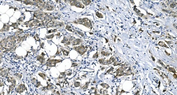
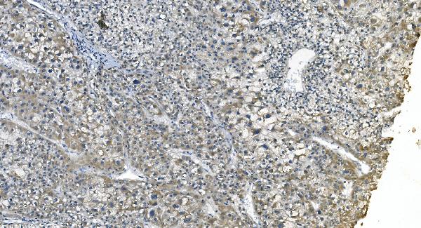
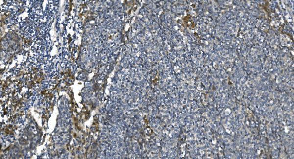
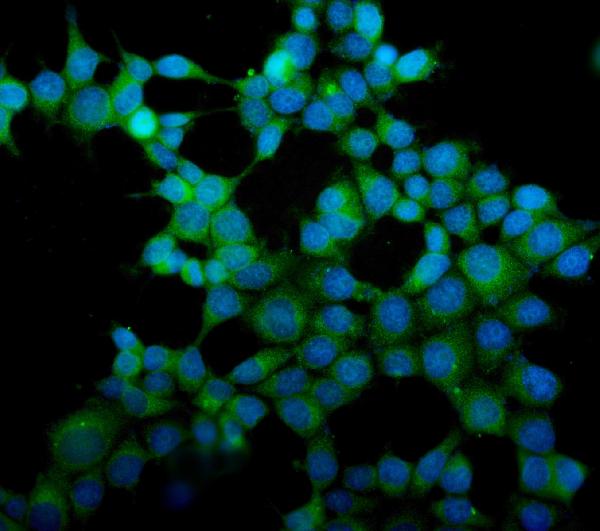
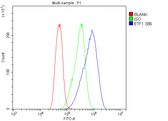
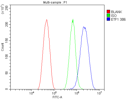
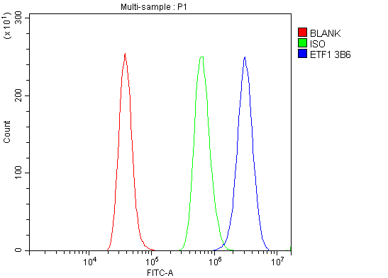
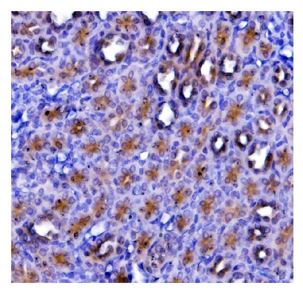
Anti-eRF1/ETF1 Antibody Picoband™ (monoclonal, 3B6)
- SPECIFICATION
- CITATIONS
- PROTOCOLS
- BACKGROUND

Application
| WB, IHC, IF, ICC, FC |
|---|---|
| Primary Accession | P62495 |
| Host | Mouse |
| Isotype | Mouse IgG2a |
| Reactivity | Rat, Human, Mouse |
| Clonality | Monoclonal |
| Format | Lyophilized |
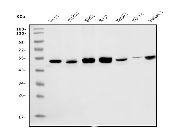
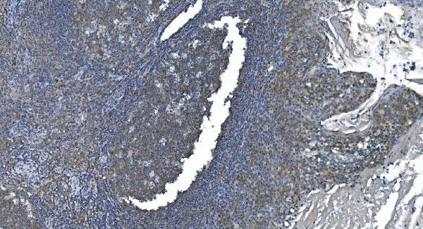
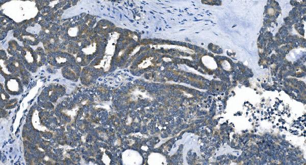
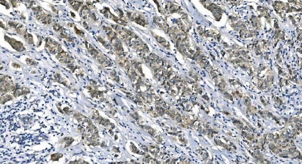
| Description | Anti-eRF1/ETF1 Antibody Picoband™ (monoclonal, 3B6) . Tested in Flow Cytometry, IF, IHC, ICC, WB applications. This antibody reacts with Human, Mouse, Rat. |
| Reconstitution | Add 0.2ml of distilled water will yield a concentration of 500ug/ml. |
| Gene ID | 2107 |
|---|---|
| Other Names | Eukaryotic peptide chain release factor subunit 1, Eukaryotic release factor 1, eRF1, Protein Cl1, TB3-1, ETF1, ERF1, RF1, SUP45L1 |
| Calculated MW | 49 kDa |
| Application Details | Western blot, 0.25-0.5 µg/ml, Human, Mouse, Rat Immunohistochemistry (Paraffin-embedded Section), 2-5 µg/ml, Human, Rat Immunocytochemistry/Immunofluorescence, 5 µg/ml, Human Flow Cytometry, 1-3 µg/1x10^6 cells, Human, Mouse, Rat |
| Contents | Each vial contains 4mg Trehalose, 0.9mg NaCl and 0.2mg Na2HPO4. |
| Clone Names | Clone: 3B6 |
| Immunogen | E.coli-derived human eRF1/ETF1 recombinant protein (Position: D9-K342). |
| Purification | Immunogen affinity purified. |
| Storage | Store at -20˚C for one year from date of receipt. After reconstitution, at 4˚C for one month. It can also be aliquotted and stored frozen at -20˚C for six months. Avoid repeated freeze-thaw cycles. |
| Name | ETF1 |
|---|---|
| Synonyms | ERF1, RF1, SUP45L1 |
| Function | Component of the eRF1-eRF3-GTP ternary complex, a ternary complex that mediates translation termination in response to the termination codons (PubMed:10676813, PubMed:16777602, PubMed:24486019, PubMed:26245381, PubMed:27863242, PubMed:36638793, PubMed:7990965). The eRF1-eRF3-GTP complex binds to a stop codon in the ribosomal A-site (PubMed:26245381, PubMed:27863242, PubMed:36638793). ETF1/ERF1 is responsible for stop codon recognition and inducing hydrolysis of peptidyl-tRNA (PubMed:26245381, PubMed:27863242, PubMed:36638793). Following GTP hydrolysis, eRF3 (GSPT1/ERF3A or GSPT2/ERF3B) dissociates, permitting ETF1/eRF1 to accommodate fully in the A-site and mediate hydrolysis of peptidyl-tRNA (PubMed:10676813, PubMed:16777602, PubMed:26245381, PubMed:27863242). Component of the transient SURF complex which recruits UPF1 to stalled ribosomes in the context of nonsense-mediated decay (NMD) of mRNAs containing premature stop codons (PubMed:19417104). Required for SHFL-mediated translation termination which inhibits programmed ribosomal frameshifting (-1PRF) of mRNA from viruses and cellular genes (PubMed:30682371). |
| Cellular Location | Cytoplasm. |

Thousands of laboratories across the world have published research that depended on the performance of antibodies from Abcepta to advance their research. Check out links to articles that cite our products in major peer-reviewed journals, organized by research category.
info@abcepta.com, and receive a free "I Love Antibodies" mug.

Provided below are standard protocols that you may find useful for product applications.
Background
Eukaryotic translation termination factor 1 (eRF1), also known asTB3-1, is a protein that in humans is encoded by the ETF1 gene. It is mapped to 5q31.2. This gene encodes a class-1 polypeptide chain release factor. The encoded protein plays an essential role in directing termination of mRNA translation from the termination codons UAA, UAG and UGA. This protein is a component of the SURF complex which promotes degradation of prematurely terminated mRNAs via the mechanism of nonsense-mediated mRNA decay (NMD). Alternate splicing results in multiple transcript variants. Pseudogenes of this gene are found on chromosomes 6, 7, and X.
If you have used an Abcepta product and would like to share how it has performed, please click on the "Submit Review" button and provide the requested information. Our staff will examine and post your review and contact you if needed.
If you have any additional inquiries please email technical services at tech@abcepta.com.
Ordering Information
Other Products
Shipping Information
