


 Foundational characteristics of cancer include proliferation, angiogenesis, migration, evasion of apoptosis, and cellular immortality. Find key markers for these cellular processes and antibodies to detect them.
 The SUMOplot™ Analysis Program predicts and scores sumoylation sites in your protein. SUMOylation is a post-translational modification involved in various cellular processes, such as nuclear-cytosolic transport, transcriptional regulation, apoptosis, protein stability, response to stress, and progression through the cell cycle.
 The Autophagy Receptor Motif Plotter predicts and scores autophagy receptor binding sites in your protein. Identifying proteins connected to this pathway is critical to understanding the role of autophagy in physiological as well as pathological processes such as development, differentiation, neurodegenerative diseases, stress, infection, and cancer.
Anti-IDH2 Antibody Picoband™ (monoclonal, 2H4)

- SPECIFICATION
- CITATIONS
- PROTOCOLS
- BACKGROUND

Application
| WB, IHC |
|---|---|
| Primary Accession | P48735 |
| Host | Mouse |
| Isotype | Mouse IgG1 |
| Reactivity | Rat, Human, Mouse |
| Clonality | Monoclonal |
| Format | Lyophilized |
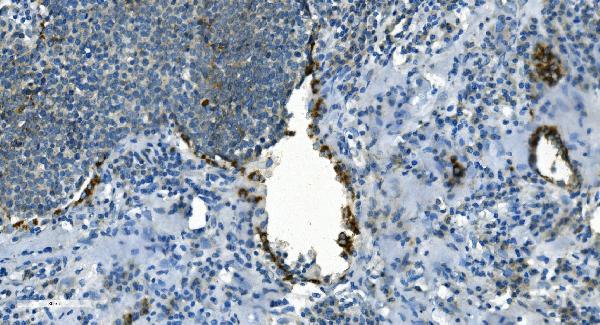
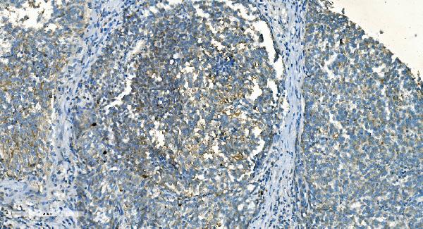
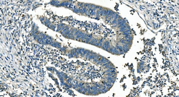
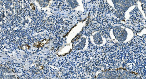
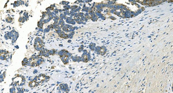
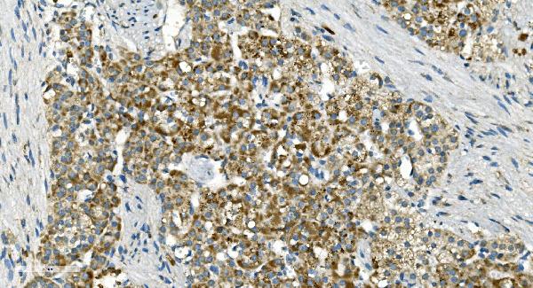
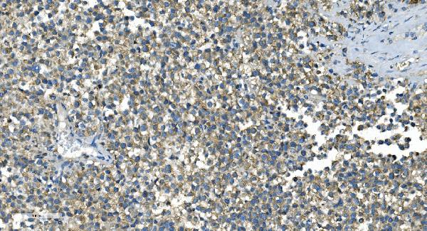
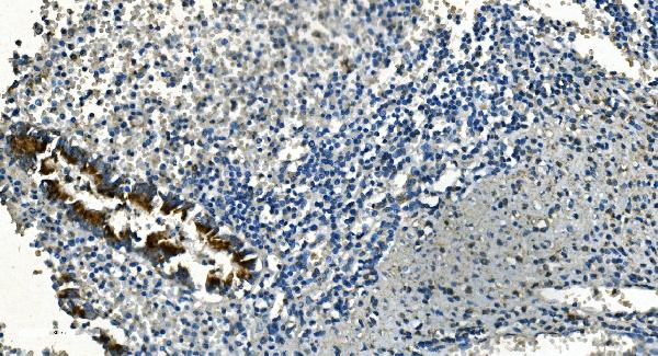
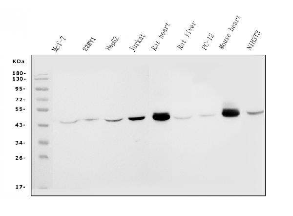
| Description | Anti-IDH2 Antibody Picoband™ (monoclonal, 2H4) . Tested in IHC, WB applications. This antibody reacts with Human, Mouse, Rat. |
| Reconstitution | Add 0.2ml of distilled water will yield a concentration of 500ug/ml. |
| Gene ID | 3418 |
|---|---|
| Other Names | Isocitrate dehydrogenase [NADP], mitochondrial, IDH, 1.1.1.42, ICD-M, IDP, NADP(+)-specific ICDH, Oxalosuccinate decarboxylase, IDH2 |
| Calculated MW | 45 kDa |
| Application Details | Western blot, 0.25-0.5 µg/ml, Human, Mouse, Rat Immunohistochemistry (Paraffin-embedded Section), 2-5 µg/ml, Human |
| Contents | Each vial contains 4mg Trehalose, 0.9mg NaCl and 0.2mg Na2HPO4. |
| Clone Names | Clone: 2H4 |
| Immunogen | A synthetic peptide corresponding to a sequence at the C-terminus of human IDH2, identical to the related mouse and rat sequences. |
| Purification | Immunogen affinity purified. |
| Storage | Store at -20˚C for one year from date of receipt. After reconstitution, at 4˚C for one month. It can also be aliquotted and stored frozen at -20˚C for six months. Avoid repeated freeze-thaw cycles. |
| Name | IDH2 |
|---|---|
| Function | Plays a role in intermediary metabolism and energy production (PubMed:19228619, PubMed:22416140). It may tightly associate or interact with the pyruvate dehydrogenase complex (PubMed:19228619, PubMed:22416140). |
| Cellular Location | Mitochondrion {ECO:0000250|UniProtKB:P33198}. |

Thousands of laboratories across the world have published research that depended on the performance of antibodies from Abcepta to advance their research. Check out links to articles that cite our products in major peer-reviewed journals, organized by research category.
info@abcepta.com, and receive a free "I Love Antibodies" mug.

Provided below are standard protocols that you may find useful for product applications.
Background
Isocitrate dehydrogenase [NADP], mitochondrialis anenzymethat in humans is encoded by theIDH2gene. Isocitrate dehydrogenases catalyze the oxidative decarboxylation of isocitrate to 2-oxoglutarate. These enzymes belong to two distinct subclasses, one of which utilizes NAD (+) as the electron acceptor and the other NADP (+). Five isocitrate dehydrogenases have been reported: three NAD (+)-dependent isocitrate dehydrogenases, which localize to the mitochondrial matrix, and two NADP (+)-dependent isocitrate dehydrogenases, one of which is mitochondrial and the other predominantly cytosolic. Each NADP (+)-dependent isozyme is a homodimer. The protein encoded by this gene is the NADP (+)-dependent isocitrate dehydrogenase found in the mitochondria. It plays a role in intermediary metabolism and energy production. This protein may tightly associate or interact with the pyruvate dehydrogenase complex. Alternative splicing results in multiple transcript variants.
If you have used an Abcepta product and would like to share how it has performed, please click on the "Submit Review" button and provide the requested information. Our staff will examine and post your review and contact you if needed.
If you have any additional inquiries please email technical services at tech@abcepta.com.
Ordering Information
Other Products
Shipping Information
