


 Foundational characteristics of cancer include proliferation, angiogenesis, migration, evasion of apoptosis, and cellular immortality. Find key markers for these cellular processes and antibodies to detect them.
 The SUMOplot™ Analysis Program predicts and scores sumoylation sites in your protein. SUMOylation is a post-translational modification involved in various cellular processes, such as nuclear-cytosolic transport, transcriptional regulation, apoptosis, protein stability, response to stress, and progression through the cell cycle.
 The Autophagy Receptor Motif Plotter predicts and scores autophagy receptor binding sites in your protein. Identifying proteins connected to this pathway is critical to understanding the role of autophagy in physiological as well as pathological processes such as development, differentiation, neurodegenerative diseases, stress, infection, and cancer.
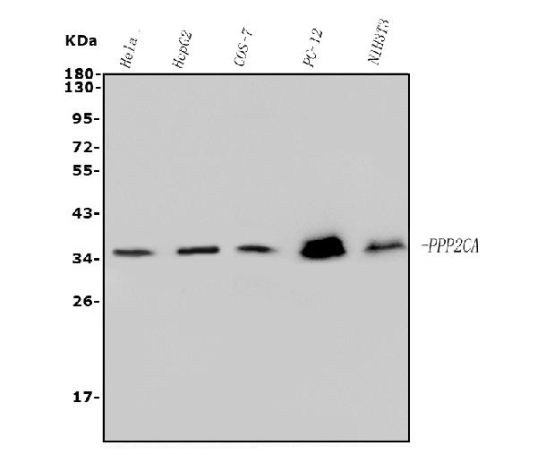
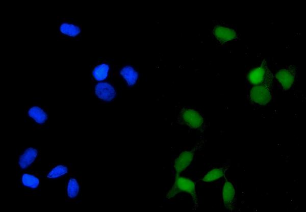
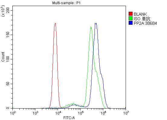
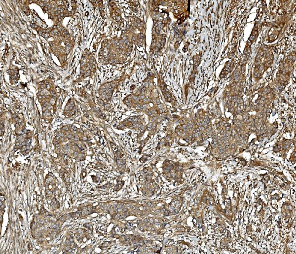
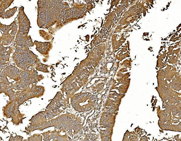
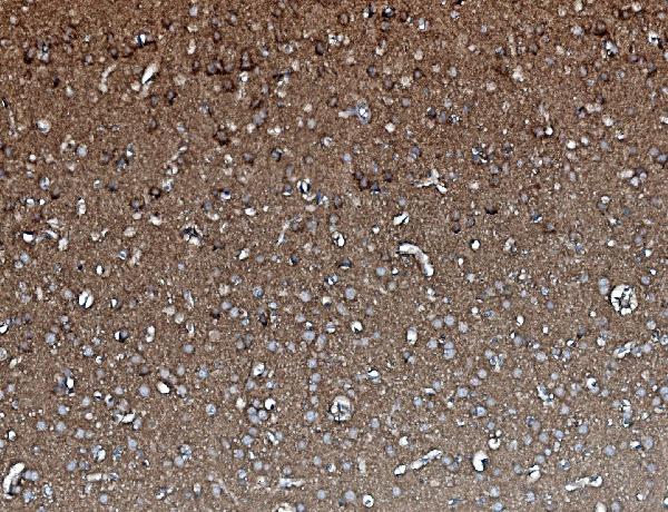
Anti-PP2A-alpha/PPP2CA Antibody Picoband™ (monoclonal, 3B6)
- SPECIFICATION
- CITATIONS
- PROTOCOLS
- BACKGROUND

Application
| WB, IHC, IF, ICC, FC |
|---|---|
| Primary Accession | P67775 |
| Host | Mouse |
| Isotype | Mouse IgG2a |
| Reactivity | Rat, Human, Mouse, Monkey |
| Clonality | Monoclonal |
| Format | Lyophilized |
| Description | Anti-PP2A-alpha/PPP2CA Antibody Picoband™ (monoclonal, 3B6) . Tested in Flow Cytometry, IF, IHC, ICC, WB applications. This antibody reacts with Human, Monkey, Mouse, Rat. |
| Reconstitution | Add 0.2ml of distilled water will yield a concentration of 500ug/ml. |
| Gene ID | 5515 |
|---|---|
| Other Names | Serine/threonine-protein phosphatase 2A catalytic subunit alpha isoform, PP2A-alpha, 3.1.3.16, Replication protein C, RP-C, PPP2CA |
| Calculated MW | 36 kDa |
| Application Details | Western blot, 0.1-0.5 µg/ml, Human, Monkey, Mouse, Rat Immunohistochemistry (Paraffin-embedded Section), 0.5-1 µg/ml, Human, Rat Immunocytochemistry/Immunofluorescence, 4 µg/ml, Human Flow Cytometry, 1-3 µg/1x10^6 cells, Human |
| Contents | Each vial contains 4mg Trehalose, 0.9mg NaCl, 0.2mg Na2HPO4, 0.01mg NaN3. |
| Clone Names | Clone: 3B6 |
| Immunogen | E.coli-derived human PP2A-alpha recombinant protein (Position: M1-L309). Human PP2A-alpha shares 100% amino acid (aa) sequence identity with both mouse and rat PP2A-alpha. |
| Purification | Immunogen affinity purified. |
| Storage | Store at -20˚C for one year from date of receipt. After reconstitution, at 4˚C for one month. It can also be aliquotted and stored frozen at -20˚C for six months. Avoid repeated freeze-thaw cycles. |
| Name | PPP2CA |
|---|---|
| Function | Catalytic subunit of protein phosphatase 2A (PP2A), a serine/threonine phosphatase involved in the regulation of a wide variety of enzymes, signal transduction pathways, and cellular events. PP2A is the major phosphatase for microtubule-associated proteins (MAPs) (PubMed:22613722). PP2A can modulate the activity of phosphorylase B kinase casein kinase 2, mitogen-stimulated S6 kinase, and MAP-2 kinase (PubMed:22613722). Cooperates with SGO2 to protect centromeric cohesin from separase-mediated cleavage in oocytes specifically during meiosis I (By similarity). Can dephosphorylate SV40 large T antigen and p53/TP53 (PubMed:17245430). Activates RAF1 by dephosphorylating it at 'Ser-259' (PubMed:10801873). Mediates dephosphorylation of WEE1, preventing its ubiquitin-mediated proteolysis, increasing WEE1 protein levels, and promoting the G2/M checkpoint (PubMed:33108758). Mediates dephosphorylation of MYC; promoting its ubiquitin-mediated proteolysis: interaction with AMBRA1 enhances interaction between PPP2CA and MYC (PubMed:25438055). Mediates dephosphorylation of FOXO3; promoting its stabilization: interaction with AMBRA1 enhances interaction between PPP2CA and FOXO3 (PubMed:30513302). Catalyzes dephosphorylation of the pyrin domain of NLRP3, promoting assembly of the NLRP3 inflammasome (By similarity). Part of the striatin-interacting phosphatase and kinase (STRIPAK) complexes. STRIPAK complexes have critical roles in protein (de)phosphorylation and are regulators of multiple signaling pathways including Hippo, MAPK, nuclear receptor and cytoskeleton remodeling. Different types of STRIPAK complexes are involved in a variety of biological processes such as cell growth, differentiation, apoptosis, metabolism and immune regulation (PubMed:33633399). |
| Cellular Location | Cytoplasm. Nucleus. Chromosome, centromere. Cytoplasm, cytoskeleton, spindle pole. Note=In prometaphase cells, but not in anaphase cells, localizes at centromeres (PubMed:16541025). During mitosis, also found at spindle poles (PubMed:16541025). Centromeric localization requires the presence of SGO2 (By similarity) {ECO:0000250|UniProtKB:P63330, ECO:0000269|PubMed:16541025} |

Thousands of laboratories across the world have published research that depended on the performance of antibodies from Abcepta to advance their research. Check out links to articles that cite our products in major peer-reviewed journals, organized by research category.
info@abcepta.com, and receive a free "I Love Antibodies" mug.

Provided below are standard protocols that you may find useful for product applications.
Background
The catalytic subunit of human protein phosphatase 2A (PPP2CA) encodes a 309-amino acid polypeptide. It is localized to chromosome 5. The gene (approximately 30 kbp) is composed of seven exons and six introns. It is predicted to be important for phosphatase enzymatic activity. Methylation of the C-terminal leucine residue (Leu309) of protein serine/threonine phosphatase 2A catalytic subunit (PP2AC) is known to regulate catalytic activity in vitro. Furthermore, PP2A has a fundamental role in cardiac function, and suggests that disturbances in protein phosphatase expression and activity may cause or exacerbate the course of cardiac diseases.
If you have used an Abcepta product and would like to share how it has performed, please click on the "Submit Review" button and provide the requested information. Our staff will examine and post your review and contact you if needed.
If you have any additional inquiries please email technical services at tech@abcepta.com.
Ordering Information
Other Products
Shipping Information
