


 Foundational characteristics of cancer include proliferation, angiogenesis, migration, evasion of apoptosis, and cellular immortality. Find key markers for these cellular processes and antibodies to detect them.
 The SUMOplot™ Analysis Program predicts and scores sumoylation sites in your protein. SUMOylation is a post-translational modification involved in various cellular processes, such as nuclear-cytosolic transport, transcriptional regulation, apoptosis, protein stability, response to stress, and progression through the cell cycle.
 The Autophagy Receptor Motif Plotter predicts and scores autophagy receptor binding sites in your protein. Identifying proteins connected to this pathway is critical to understanding the role of autophagy in physiological as well as pathological processes such as development, differentiation, neurodegenerative diseases, stress, infection, and cancer.
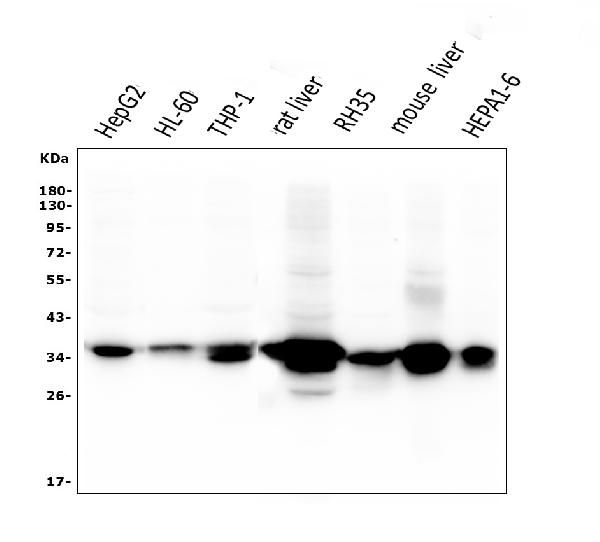
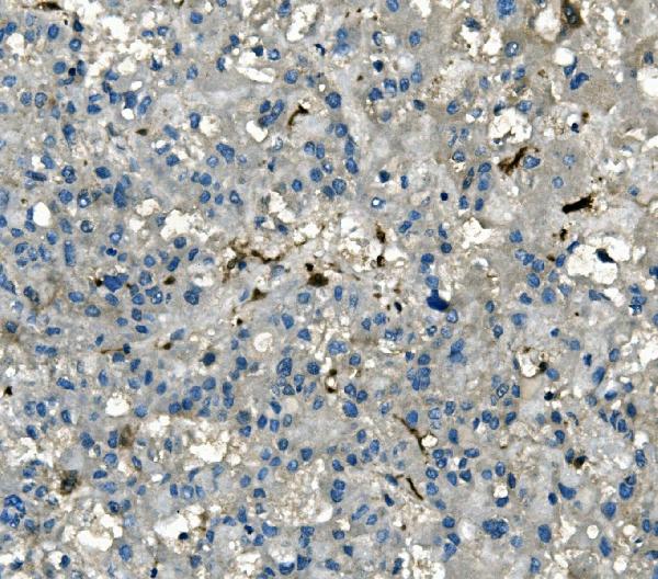
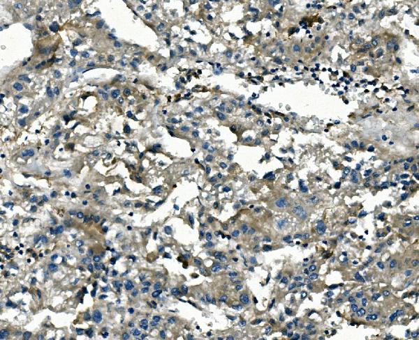
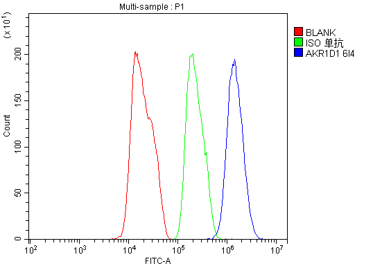
Anti-AKR1D1 Antibody Picoband™ (monoclonal, 6I4)
- SPECIFICATION
- CITATIONS
- PROTOCOLS
- BACKGROUND

Application
| WB, IHC, FC |
|---|---|
| Primary Accession | P51857 |
| Host | Mouse |
| Isotype | Mouse IgG2b |
| Reactivity | Rat, Human, Mouse |
| Clonality | Monoclonal |
| Format | Lyophilized |
| Description | Anti-AKR1D1 Antibody Picoband™ (monoclonal, 6I4) . Tested in Flow Cytometry, IHC, WB applications. This antibody reacts with Human, Mouse, Rat. |
| Reconstitution | Add 0.2ml of distilled water will yield a concentration of 500ug/ml. |
| Gene ID | 6718 |
|---|---|
| Other Names | Aldo-keto reductase family 1 member D1, 1.3.1.3, 3-oxo-5-beta-steroid 4-dehydrogenase, Delta(4)-3-ketosteroid 5-beta-reductase, Delta(4)-3-oxosteroid 5-beta-reductase, AKR1D1, SRD5B1 |
| Calculated MW | 37 kDa |
| Application Details | Western blot, 0.1-0.5 µg/ml, Human, Mouse, Rat Immunohistochemistry (Paraffin-embedded Section), 0.5-1 µg/ml, Human Flow Cytometry, 1-3 µg/1x10^6 cells, Human |
| Subcellular Localization | Cytoplasm. |
| Tissue Specificity | Highly expressed in liver. Expressed in testis and weakly in colon. |
| Contents | Each vial contains 4mg Trehalose, 0.9mg NaCl, 0.2mg Na2HPO4, 0.05mg NaN3. |
| Clone Names | Clone: 6I4 |
| Immunogen | A synthetic peptide corresponding to a sequence at the C-terminus of human AKR1D1, which shares 90.9% and 93.9% amino acid (aa) sequence identity with mouse and rat AKR1D1, respectively. |
| Purification | Immunogen affinity purified. |
| Cross Reactivity | No cross-reactivity with other proteins. |
| Storage | Store at -20˚C for one year from date of receipt. After reconstitution, at 4˚C for one month. It can also be aliquotted and stored frozen at -20˚C for six months. Avoid repeated freeze-thaw cycles. |
| Name | AKR1D1 |
|---|---|
| Synonyms | SRD5B1 |
| Function | Catalyzes the stereospecific NADPH-dependent reduction of the C4-C5 double bond of bile acid intermediates and steroid hormones carrying a delta(4)-3-one structure to yield an A/B cis-ring junction. This cis-configuration is crucial for bile acid biosynthesis and plays important roles in steroid metabolism. Capable of reducing a broad range of delta-(4)-3-ketosteroids from C18 (such as, 17beta- hydroxyestr-4-en-3-one) to C27 (such as, 7alpha-hydroxycholest-4-en-3- one). |
| Cellular Location | Cytoplasm. |
| Tissue Location | Highly expressed in liver. Expressed in testis and weakly in colon. |

Thousands of laboratories across the world have published research that depended on the performance of antibodies from Abcepta to advance their research. Check out links to articles that cite our products in major peer-reviewed journals, organized by research category.
info@abcepta.com, and receive a free "I Love Antibodies" mug.

Provided below are standard protocols that you may find useful for product applications.
Background
Human delta (4)-3-oxosteroid 5-beta-reductase (steroid 5-beta-reductase) catalyzes 5-beta-reduction of bile acid intermediates and steroid hormones carrying a delta (4)-3-one structure. This gene is mapped to 7q33. The enzyme encoded by this gene is responsible for the catalysis of the 5-beta-reduction of bile acid intermediates and steroid hormones carrying a delta (4)-3-one structure. Deficiency of this enzyme may contribute to hepatic dysfunction. Three transcript variants encoding different isoforms have been found for this gene. Other variants may be present, but their full-length natures have not been determined yet.
If you have used an Abcepta product and would like to share how it has performed, please click on the "Submit Review" button and provide the requested information. Our staff will examine and post your review and contact you if needed.
If you have any additional inquiries please email technical services at tech@abcepta.com.
Ordering Information
Other Products
Shipping Information
