


 Foundational characteristics of cancer include proliferation, angiogenesis, migration, evasion of apoptosis, and cellular immortality. Find key markers for these cellular processes and antibodies to detect them.
 The SUMOplot™ Analysis Program predicts and scores sumoylation sites in your protein. SUMOylation is a post-translational modification involved in various cellular processes, such as nuclear-cytosolic transport, transcriptional regulation, apoptosis, protein stability, response to stress, and progression through the cell cycle.
 The Autophagy Receptor Motif Plotter predicts and scores autophagy receptor binding sites in your protein. Identifying proteins connected to this pathway is critical to understanding the role of autophagy in physiological as well as pathological processes such as development, differentiation, neurodegenerative diseases, stress, infection, and cancer.
> home > Products > Primary Antibodies > Signal Transduction > Anti-ZAP70 Antibody Picoband™ (monoclonal, 9D5)
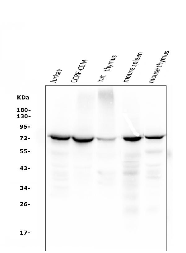
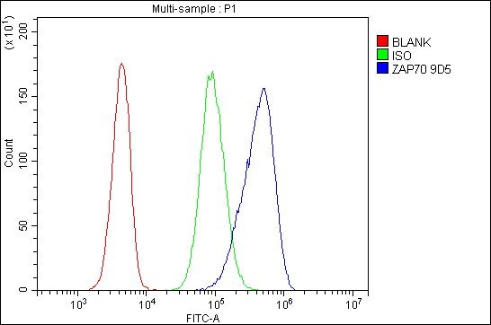
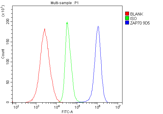
Anti-ZAP70 Antibody Picoband™ (monoclonal, 9D5)
- SPECIFICATION
- CITATIONS
- PROTOCOLS
- BACKGROUND

Application
| WB, FC |
|---|---|
| Primary Accession | P43403 |
| Host | Mouse |
| Isotype | Mouse IgG2b |
| Reactivity | Rat, Human, Mouse |
| Clonality | Monoclonal |
| Format | Lyophilized |
| Description | Anti-ZAP70 Antibody Picoband™ (monoclonal, 9D5) . Tested in Flow Cytometry, WB applications. This antibody reacts with Human, Mouse, Rat. |
| Reconstitution | Add 0.2ml of distilled water will yield a concentration of 500ug/ml. |
| Gene ID | 7535 |
|---|---|
| Other Names | Tyrosine-protein kinase ZAP-70, 2.7.10.2, 70 kDa zeta-chain associated protein, Syk-related tyrosine kinase, ZAP70, SRK |
| Calculated MW | 72 kDa |
| Application Details | Western blot, 0.1-0.5 µg/ml, Human, Mouse, Rat Flow Cytometry, 1-3 µg/1x10^6 cells, Human |
| Subcellular Localization | Cell membrane. Cytoplasm. Membrane. |
| Tissue Specificity | Expressed in T- and natural killer cells. Also present in early thymocytes and pro/pre B-cells. |
| Contents | Each vial contains 4mg Trehalose, 0.9mg NaCl, 0.2mg Na2HPO4, 0.05mg NaN3. |
| Clone Names | Clone: 9D5 |
| Immunogen | A synthetic peptide corresponding to a sequence in the middle region of human ZAP70, different from the related mouse sequence by two amino acids. |
| Purification | Immunogen affinity purified. |
| Cross Reactivity | No cross-reactivity with other proteins. |
| Storage | Store at -20˚C for one year from date of receipt. After reconstitution, at 4˚C for one month. It can also be aliquotted and stored frozen at -20˚C for six months. Avoid repeated freeze-thaw cycles. |
| Name | ZAP70 |
|---|---|
| Synonyms | SRK |
| Function | Tyrosine kinase that plays an essential role in regulation of the adaptive immune response. Regulates motility, adhesion and cytokine expression of mature T-cells, as well as thymocyte development. Contributes also to the development and activation of primary B- lymphocytes. When antigen presenting cells (APC) activate T-cell receptor (TCR), a serie of phosphorylations lead to the recruitment of ZAP70 to the doubly phosphorylated TCR component CD247/CD3Z through ITAM motif at the plasma membrane. This recruitment serves to localization to the stimulated TCR and to relieve its autoinhibited conformation. Release of ZAP70 active conformation is further stabilized by phosphorylation mediated by LCK. Subsequently, ZAP70 phosphorylates at least 2 essential adapter proteins: LAT and LCP2. In turn, a large number of signaling molecules are recruited and ultimately lead to lymphokine production, T-cell proliferation and differentiation. Furthermore, ZAP70 controls cytoskeleton modifications, adhesion and mobility of T-lymphocytes, thus ensuring correct delivery of effectors to the APC. ZAP70 is also required for TCR-CD247/CD3Z internalization and degradation through interaction with the E3 ubiquitin-protein ligase CBL and adapter proteins SLA and SLA2. Thus, ZAP70 regulates both T-cell activation switch on and switch off by modulating TCR expression at the T-cell surface. During thymocyte development, ZAP70 promotes survival and cell-cycle progression of developing thymocytes before positive selection (when cells are still CD4/CD8 double negative). Additionally, ZAP70-dependent signaling pathway may also contribute to primary B-cells formation and activation through B-cell receptor (BCR). |
| Cellular Location | Cytoplasm. Cell membrane; Peripheral membrane protein. Note=In quiescent T-lymphocytes, it is cytoplasmic. Upon TCR activation, it is recruited at the plasma membrane by interacting with CD247/CD3Z. Colocalizes together with RHOH in the immunological synapse. RHOH is required for its proper localization to the cell membrane and cytoskeleton fractions in the thymocytes (By similarity). |
| Tissue Location | Expressed in T- and natural killer cells. Also present in early thymocytes and pro/pre B-cells |
Research Areas

Abcepta welcomes feedback from its customers.
If you have used an Abcepta product and would like to share how it has performed, please click on the "Submit Review" button and provide the requested information. Our staff will examine and post your review and contact you if needed.
If you have any additional inquiries please email technical services at tech@abcepta.com.
$ 370.00
Cat# ABO14951
Ordering Information
United States
AlbaniaAustraliaAustriaBelgiumBosnia & HerzegovinaBrazilBulgariaCanadaCentral AmericaChinaCroatiaCyprusCzech RepublicDenmarkEstoniaFinlandFranceGermanyGreeceHong KongHungaryIcelandIndiaIndonesiaIrelandIsraelItalyJapanLatviaLithuaniaLuxembourgMacedoniaMalaysiaMaltaNetherlandsNew ZealandNorwayPakistanPolandPortugalRomaniaSerbiaSingaporeSlovakiaSloveniaSouth AfricaSouth KoreaSpainSwedenSwitzerlandTaiwanTurkeyUnited KingdomUnited StatesVietnamWorldwideOthersMexico
USA Headquarters
(888) 735-7227 / (858) 622-0099 or (858) 875-1900
Other Products
Shipping Information
Domestic orders (in stock items)
Shipped out the same day. Orders placed after 1 PM (PST) will ship out the next business day.
International orders
Contact your local distributors
