


 Foundational characteristics of cancer include proliferation, angiogenesis, migration, evasion of apoptosis, and cellular immortality. Find key markers for these cellular processes and antibodies to detect them.
 The SUMOplot™ Analysis Program predicts and scores sumoylation sites in your protein. SUMOylation is a post-translational modification involved in various cellular processes, such as nuclear-cytosolic transport, transcriptional regulation, apoptosis, protein stability, response to stress, and progression through the cell cycle.
 The Autophagy Receptor Motif Plotter predicts and scores autophagy receptor binding sites in your protein. Identifying proteins connected to this pathway is critical to understanding the role of autophagy in physiological as well as pathological processes such as development, differentiation, neurodegenerative diseases, stress, infection, and cancer.
Anti-Thioredoxin 2/TXN2 Antibody Picoband™ (monoclonal, 4H3)

- SPECIFICATION
- CITATIONS
- PROTOCOLS
- BACKGROUND

Application
| WB, IHC, IF, ICC, FC |
|---|---|
| Primary Accession | Q99757 |
| Host | Mouse |
| Isotype | Mouse IgG2a |
| Reactivity | Rat, Human, Mouse |
| Clonality | Monoclonal |
| Format | Lyophilized |
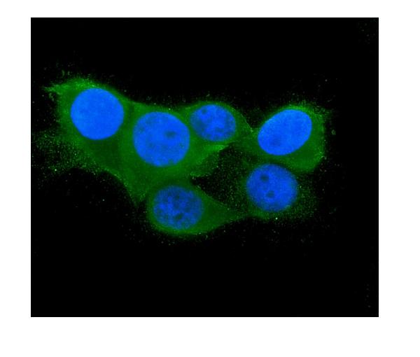
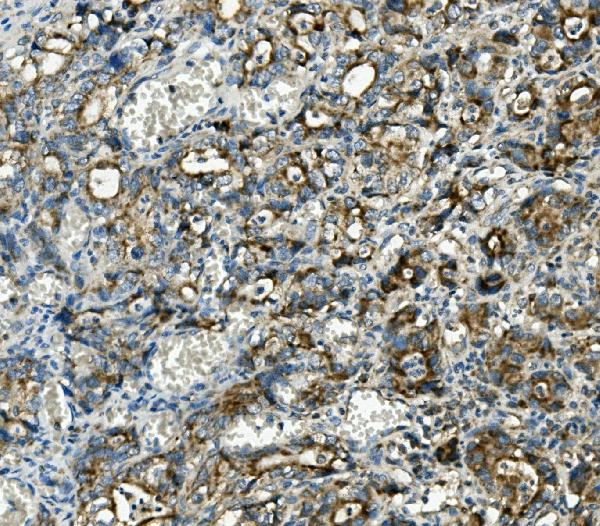
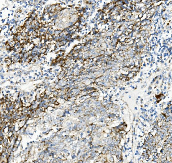
| Description | Anti-Thioredoxin 2/TXN2 Antibody Picoband™ (monoclonal, 4H3) . Tested in Flow Cytometry, IF, IHC, ICC, WB applications. This antibody reacts with Human, Mouse, Rat. |
| Reconstitution | Add 0.2ml of distilled water will yield a concentration of 500ug/ml. |
| Gene ID | 25828 |
|---|---|
| Other Names | Thioredoxin, mitochondrial, MTRX, Mt-Trx, Thioredoxin-2, TXN2, TRX2 |
| Calculated MW | 14 kDa |
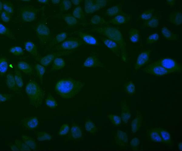
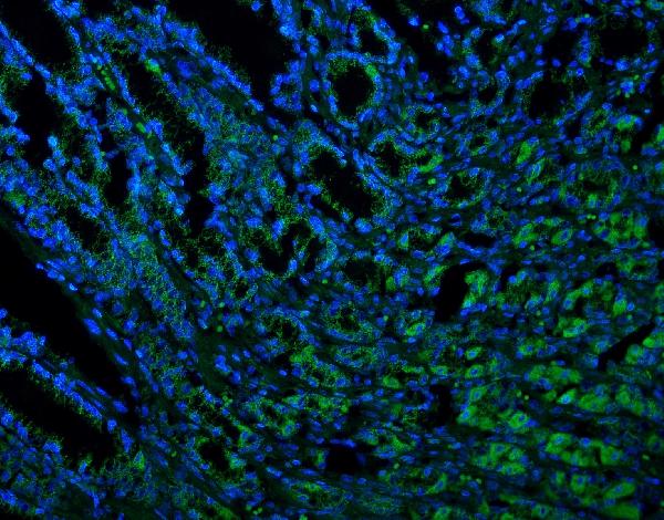
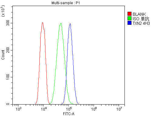
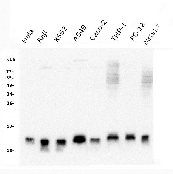
| Application Details | Western blot, 0.1-0.5 µg/ml, Human, Mouse, Rat Immunohistochemistry (Paraffin-embedded Section), 0.5-1 µg/ml, Human, Mouse, Rat Immunofluorescence, 2 µg/ml, Human Immunocytochemistry/Immunofluorescence, 2 µg/ml, Human Flow Cytometry, 1-3 µg/1x10^6 cells, Human |
| Subcellular Localization | Mitochondrion. |
| Tissue Specificity | Widely expressed in adult (at protein level) and fetal tissues. |
| Contents | Each vial contains 4mg Trehalose, 0.9mg NaCl, 0.2mg Na2HPO4, 0.05mg NaN3. |
| Clone Names | Clone: 4H3 |
| Immunogen | E.coli-derived human Thioredoxin 2/TXN2 recombinant protein (Position: T60-G166). |
| Purification | Immunogen affinity purified. |
| Cross Reactivity | No cross-reactivity with other proteins. |
| Storage | Store at -20˚C for one year from date of receipt. After reconstitution, at 4˚C for one month. It can also be aliquotted and stored frozen at -20˚C for six months. Avoid repeated freeze-thaw cycles. |
| Name | TXN2 |
|---|---|
| Synonyms | TRX2 |
| Function | Important for the control of mitochondrial reactive oxygen species homeostasis, apoptosis regulation and cell viability. Possesses a dithiol-reducing activity. |
| Cellular Location | Mitochondrion |
| Tissue Location | Widely expressed in adult (at protein level) and fetal tissues. |

Thousands of laboratories across the world have published research that depended on the performance of antibodies from Abcepta to advance their research. Check out links to articles that cite our products in major peer-reviewed journals, organized by research category.
info@abcepta.com, and receive a free "I Love Antibodies" mug.

Provided below are standard protocols that you may find useful for product applications.
Background
Thioredoxin, mitochondrial also known as thioredoxin-2 is a protein that in humans is encoded by the TXN2 gene on chromosome 22. It is mapped to 22q12.3. This nuclear gene encodes a mitochondrial member of the thioredoxin family, a group of small multifunctional redox-active proteins. The encoded protein may play important roles in the regulation of the mitochondrial membrane potential and in protection against oxidant-induced apoptosis.
If you have used an Abcepta product and would like to share how it has performed, please click on the "Submit Review" button and provide the requested information. Our staff will examine and post your review and contact you if needed.
If you have any additional inquiries please email technical services at tech@abcepta.com.
Ordering Information
Other Products
Shipping Information
