


 Foundational characteristics of cancer include proliferation, angiogenesis, migration, evasion of apoptosis, and cellular immortality. Find key markers for these cellular processes and antibodies to detect them.
 The SUMOplot™ Analysis Program predicts and scores sumoylation sites in your protein. SUMOylation is a post-translational modification involved in various cellular processes, such as nuclear-cytosolic transport, transcriptional regulation, apoptosis, protein stability, response to stress, and progression through the cell cycle.
 The Autophagy Receptor Motif Plotter predicts and scores autophagy receptor binding sites in your protein. Identifying proteins connected to this pathway is critical to understanding the role of autophagy in physiological as well as pathological processes such as development, differentiation, neurodegenerative diseases, stress, infection, and cancer.
Anti-alpha 1 Catenin/CTNNA1 Antibody Picoband™ (monoclonal, 10I2)
- SPECIFICATION
- CITATIONS
- PROTOCOLS
- BACKGROUND

Application
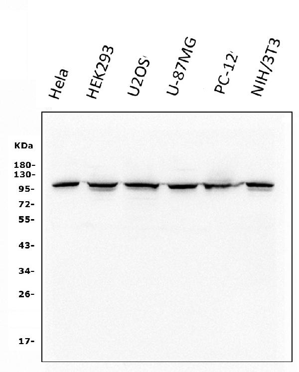
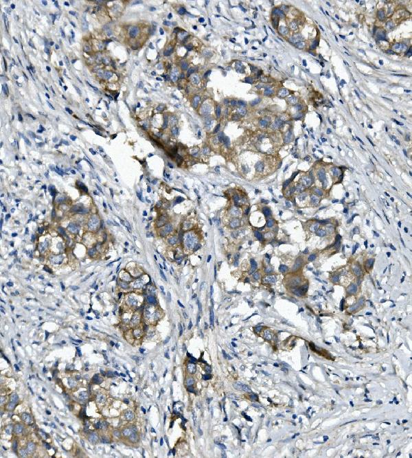
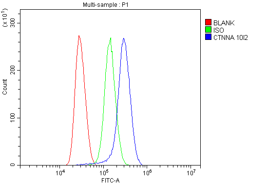
| WB, IHC, FC |
|---|---|
| Primary Accession | P35221 |
| Host | Mouse |
| Isotype | Mouse IgG1 |
| Reactivity | Rat, Human, Mouse |
| Clonality | Monoclonal |
| Format | Lyophilized |
| Description | Anti-alpha 1 Catenin/CTNNA1 Antibody Picoband™ (monoclonal, 10I2) . Tested in Flow Cytometry, IHC, WB applications. This antibody reacts with Human, Mouse, Rat. |
| Reconstitution | Add 0.2ml of distilled water will yield a concentration of 500ug/ml. |
| Gene ID | 1495 |
|---|---|
| Other Names | Catenin alpha-1 {ECO:0000312|HGNC:HGNC:2509}, Alpha E-catenin {ECO:0000312|HGNC:HGNC:2509}, Cadherin-associated protein {ECO:0000312|HGNC:HGNC:2509}, Renal carcinoma antigen NY-REN-13, CTNNA1 (HGNC:2509) |
| Calculated MW | 100 kDa |
| Application Details | Western blot, 0.1-0.5 µg/ml, Human, Mouse, Rat Immunohistochemistry (Paraffin-embedded Section), 0.5-1 µg/ml, Human Flow Cytometry, 1-3 µg/1x10^6 cells, Human |
| Subcellular Localization | Cytoskeleton. Cell membrane. Peripheral membrane protein. Cytoplasmic side. Adherens junction. Cell junction. |
| Tissue Specificity | Expressed ubiquitously in normal tissues. |
| Contents | Each vial contains 4mg Trehalose, 0.9mg NaCl, 0.2mg Na2HPO4, 0.05mg NaN3. |
| Clone Names | Clone: 10I2 |
| Immunogen | E.coli-derived human CTNNA1 recombinant protein (Position: D143-D292). Human CTNNA1 shares 98% amino acid (aa) sequence identity with mouse CTNNA1. |
| Purification | Immunogen affinity purified. |
| Cross Reactivity | No cross-reactivity with other proteins. |
| Storage | Store at -20˚C for one year from date of receipt. After reconstitution, at 4˚C for one month. It can also be aliquotted and stored frozen at -20˚C for six months. Avoid repeated freeze-thaw cycles. |
| Name | CTNNA1 (HGNC:2509) |
|---|---|
| Function | Associates with the cytoplasmic domain of a variety of cadherins. The association of catenins to cadherins produces a complex which is linked to the actin filament network, and which seems to be of primary importance for cadherins cell-adhesion properties. Can associate with both E- and N-cadherins. Originally believed to be a stable component of E-cadherin/catenin adhesion complexes and to mediate the linkage of cadherins to the actin cytoskeleton at adherens junctions. In contrast, cortical actin was found to be much more dynamic than E-cadherin/catenin complexes and CTNNA1 was shown not to bind to F-actin when assembled in the complex suggesting a different linkage between actin and adherens junctions components. The homodimeric form may regulate actin filament assembly and inhibit actin branching by competing with the Arp2/3 complex for binding to actin filaments. Involved in the regulation of WWTR1/TAZ, YAP1 and TGFB1- dependent SMAD2 and SMAD3 nuclear accumulation (By similarity). May play a crucial role in cell differentiation. |
| Cellular Location | Cytoplasm, cytoskeleton {ECO:0000250|UniProtKB:P26231}. Cell junction, adherens junction. Cell membrane {ECO:0000250|UniProtKB:P26231}; Peripheral membrane protein; Cytoplasmic side {ECO:0000250|UniProtKB:P26231}. Cell junction Cytoplasm {ECO:0000250|UniProtKB:Q9PVF8}. Nucleus. Note=Found at cell-cell boundaries and probably at cell-matrix boundaries. {ECO:0000250|UniProtKB:P26231} |
| Tissue Location | [Isoform 1]: Ubiquitously expressed in normal tissues. |

Thousands of laboratories across the world have published research that depended on the performance of antibodies from Abcepta to advance their research. Check out links to articles that cite our products in major peer-reviewed journals, organized by research category.
info@abcepta.com, and receive a free "I Love Antibodies" mug.

Provided below are standard protocols that you may find useful for product applications.
Background
CTNNA1, also known as Catenin alpha-1 or Catenin (cadherin-associated protein), alpha 1, is a protein that in humans is encoded by the CTNNA1 gene. It is mapped to 5q31.2. When surface epithelium CTNNA1 was ablated, hair follicle development was blocked and epidermal morphogenesis was dramatically affected, with defects in adherens junction formation, intercellular adhesion, and epithelial polarity. In vitro, CTNNA1 null keratinocytes were poorly contact inhibited and grew rapidly. These differences were not dependent upon intercellular adhesion and were in marked contrast to keratinocytes conditionally null for another essential intercellular adhesion protein, desmoplakin Knockout keratinocytes exhibited sustained activation of the Ras-MAPK cascade due to aberrations in growth factor responses. It is concluded that features of precancerous lesions often attributed to defects in cell cycle regulatory genes can be generated by compromising the function of CTNNA1.
If you have used an Abcepta product and would like to share how it has performed, please click on the "Submit Review" button and provide the requested information. Our staff will examine and post your review and contact you if needed.
If you have any additional inquiries please email technical services at tech@abcepta.com.
Ordering Information
Other Products
Shipping Information
