


 Foundational characteristics of cancer include proliferation, angiogenesis, migration, evasion of apoptosis, and cellular immortality. Find key markers for these cellular processes and antibodies to detect them.
 The SUMOplot™ Analysis Program predicts and scores sumoylation sites in your protein. SUMOylation is a post-translational modification involved in various cellular processes, such as nuclear-cytosolic transport, transcriptional regulation, apoptosis, protein stability, response to stress, and progression through the cell cycle.
 The Autophagy Receptor Motif Plotter predicts and scores autophagy receptor binding sites in your protein. Identifying proteins connected to this pathway is critical to understanding the role of autophagy in physiological as well as pathological processes such as development, differentiation, neurodegenerative diseases, stress, infection, and cancer.
Anti-CD146/MCAM Antibody Picoband™ (monoclonal, 4C12)

- SPECIFICATION
- CITATIONS
- PROTOCOLS
- BACKGROUND

Application
| WB, IHC, IHC-F, FC |
|---|---|
| Primary Accession | P43121 |
| Host | Mouse |
| Isotype | Mouse IgG2a |
| Reactivity | Human |
| Clonality | Monoclonal |
| Format | Lyophilized |
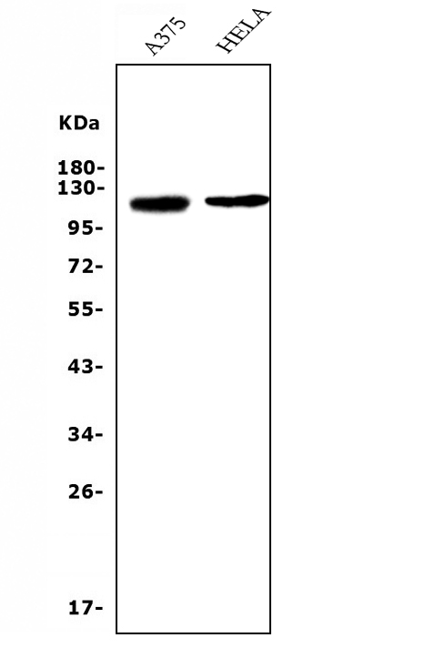
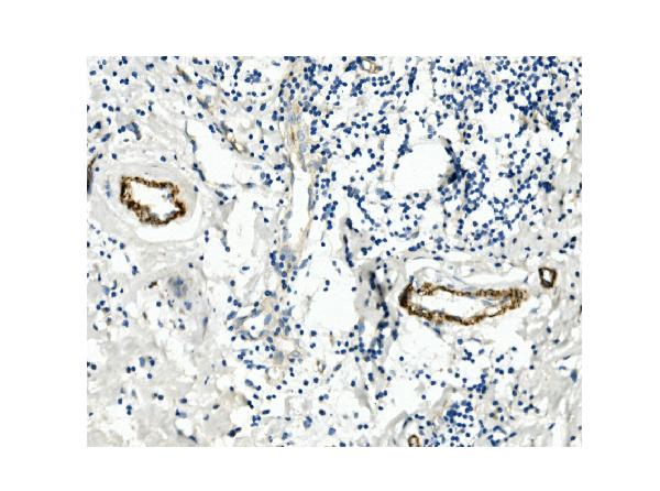
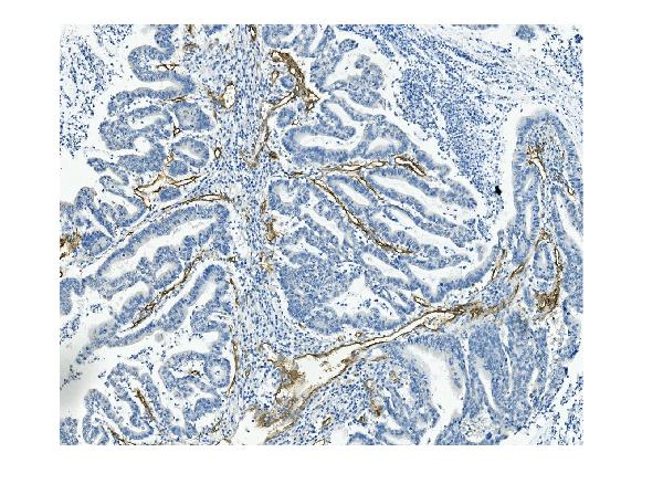
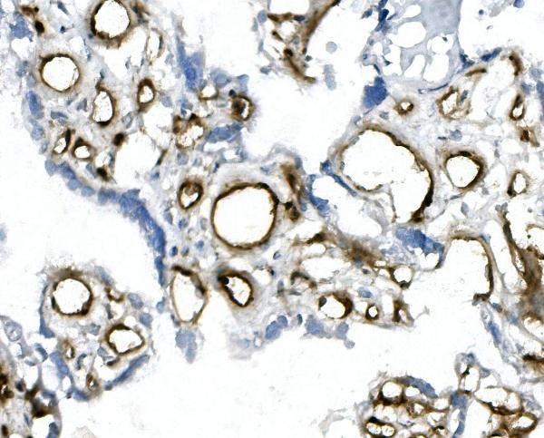
| Description | Anti-CD146/MCAM Antibody Picoband™ (monoclonal, 4C12) . Tested in Flow Cytometry, IHC, IHC-F, WB applications. This antibody reacts with Human. |
| Reconstitution | Add 0.2ml of distilled water will yield a concentration of 500ug/ml. |
| Gene ID | 4162 |
|---|---|
| Other Names | Cell surface glycoprotein MUC18, Cell surface glycoprotein P1H12, Melanoma cell adhesion molecule, Melanoma-associated antigen A32, Melanoma-associated antigen MUC18, S-endo 1 endothelial-associated antigen, CD146, MCAM, MUC18 |
| Calculated MW | 120 kDa |
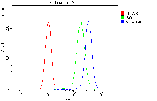
| Application Details | Western blot, 0.1-0.5 µg/ml, Human Immunohistochemistry (Paraffin-embedded Section), 0.5-1 µg/ml, Human Immunohistochemistry (Frozen Section), 0.5-1 µg/ml, Human Flow Cytometry, 1-3 µg/1x10^6 cells, Human |
| Subcellular Localization | Membrane. Single-pass type I membrane protein. |
| Tissue Specificity | Detected in endothelial cells in vascular tissue throughout the body. May appear at the surface of neural crest cells during their embryonic migration. Appears to be limited to vascular smooth muscle in normal adult tissues. Associated with tumor progression and the development of metastasis in human malignant melanoma. Expressed most strongly on metastatic lesions and advanced primary tumors and is only rarely detected in benign melanocytic nevi and thin primary melanomas with a low probability of metastasis. |
| Contents | Each vial contains 4mg Trehalose, 0.9mg NaCl, 0.2mg Na2HPO4, 0.05mg NaN3. |
| Clone Names | Clone: 4C12 |
| Immunogen | E.coli-derived human CD146 recombinant protein (Position: H59-A401). Human CD146 shares 73% amino acid (aa) sequence identity with both mouse and rat CD146. |
| Purification | Immunogen affinity purified. |
| Cross Reactivity | No cross-reactivity with other proteins. |
| Storage | Store at -20˚C for one year from date of receipt. After reconstitution, at 4˚C for one month. It can also be aliquotted and stored frozen at -20˚C for six months. Avoid repeated freeze-thaw cycles. |
| Name | MCAM |
|---|---|
| Synonyms | MUC18 |
| Function | Plays a role in cell adhesion, and in cohesion of the endothelial monolayer at intercellular junctions in vascular tissue. Its expression may allow melanoma cells to interact with cellular elements of the vascular system, thereby enhancing hematogeneous tumor spread. Could be an adhesion molecule active in neural crest cells during embryonic development. Acts as a surface receptor that triggers tyrosine phosphorylation of FYN and PTK2/FAK1, and a transient increase in the intracellular calcium concentration. |
| Cellular Location | Membrane; Single-pass type I membrane protein. |
| Tissue Location | Detected in endothelial cells in vascular tissue throughout the body. May appear at the surface of neural crest cells during their embryonic migration. Appears to be limited to vascular smooth muscle in normal adult tissues. Associated with tumor progression and the development of metastasis in human malignant melanoma. Expressed most strongly on metastatic lesions and advanced primary tumors and is only rarely detected in benign melanocytic nevi and thin primary melanomas with a low probability of metastasis |

Thousands of laboratories across the world have published research that depended on the performance of antibodies from Abcepta to advance their research. Check out links to articles that cite our products in major peer-reviewed journals, organized by research category.
info@abcepta.com, and receive a free "I Love Antibodies" mug.

Provided below are standard protocols that you may find useful for product applications.
Background
CD146 (cluster of differentiation 146), also known as the melanoma cell adhesion molecule (MCAM) or cell surface glycoprotein MUC18, is a 113kDa cell adhesion molecule currently used as a marker for endothelial cell lineage. MCAM, a member of the immunoglobulin superfamily, is homologous to several cell adhesion molecules and is associated with tumor progression and the development of metastasis in human malignant melanoma. By radiation hybrid analysis, this gene is mapped to chromosome 11q23.3. MCAM has been demonstrated to appear on a small subset of T and B lymphocytes in the peripheral blood of healthy individuals. MCAM has been seen as a marker for mesenchymal stem cells isolated from multiple adult and fetal organs, and its expression may be linked to multipotency mesenchymal stem cells with greater differentiation potential express higher levels of MCAM on the cell surface.
If you have used an Abcepta product and would like to share how it has performed, please click on the "Submit Review" button and provide the requested information. Our staff will examine and post your review and contact you if needed.
If you have any additional inquiries please email technical services at tech@abcepta.com.
Ordering Information
Other Products
Shipping Information
