


 Foundational characteristics of cancer include proliferation, angiogenesis, migration, evasion of apoptosis, and cellular immortality. Find key markers for these cellular processes and antibodies to detect them.
 The SUMOplot™ Analysis Program predicts and scores sumoylation sites in your protein. SUMOylation is a post-translational modification involved in various cellular processes, such as nuclear-cytosolic transport, transcriptional regulation, apoptosis, protein stability, response to stress, and progression through the cell cycle.
 The Autophagy Receptor Motif Plotter predicts and scores autophagy receptor binding sites in your protein. Identifying proteins connected to this pathway is critical to understanding the role of autophagy in physiological as well as pathological processes such as development, differentiation, neurodegenerative diseases, stress, infection, and cancer.
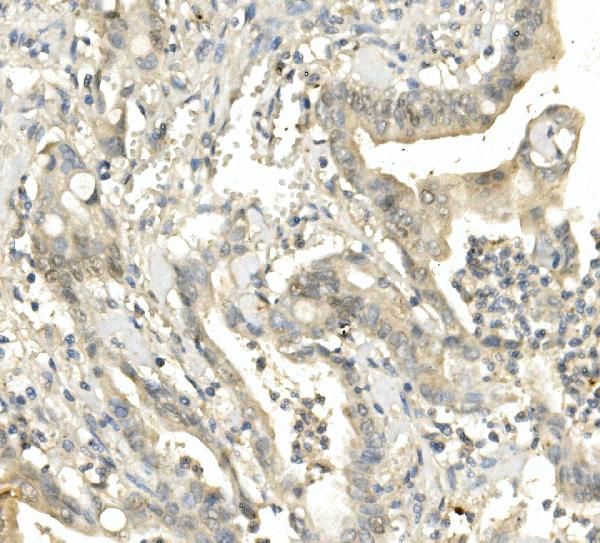
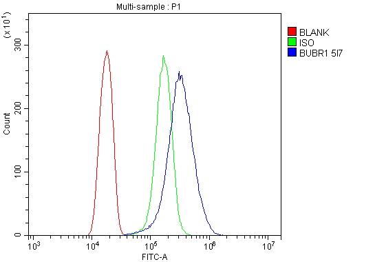
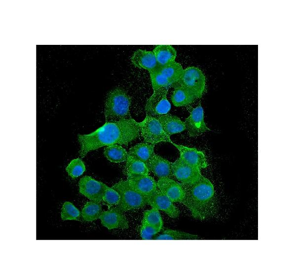
Anti-BubR1/BUB1B Antibody Picoband™ (monoclonal, 5I7)
- SPECIFICATION
- CITATIONS
- PROTOCOLS
- BACKGROUND

Application
| WB, IHC, IF, ICC, FC |
|---|---|
| Primary Accession | O60566 |
| Host | Mouse |
| Isotype | Mouse IgG1 |
| Reactivity | Human |
| Clonality | Monoclonal |
| Format | Lyophilized |
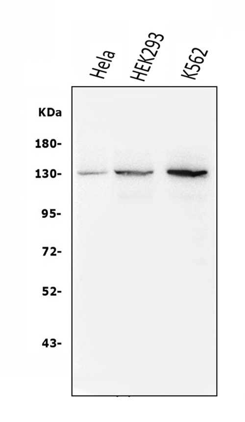
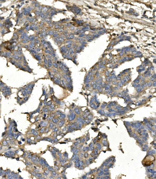
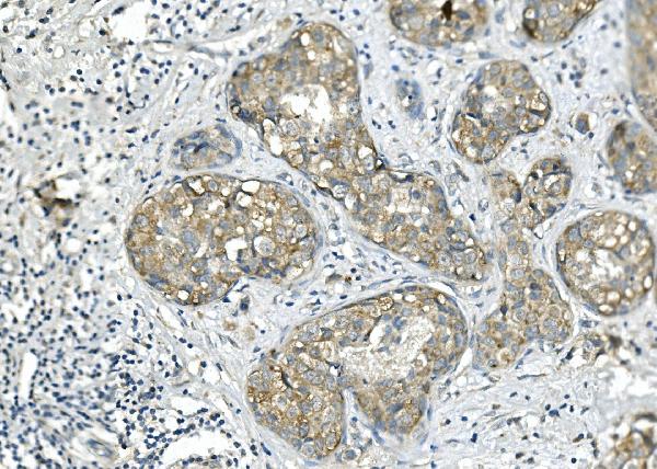
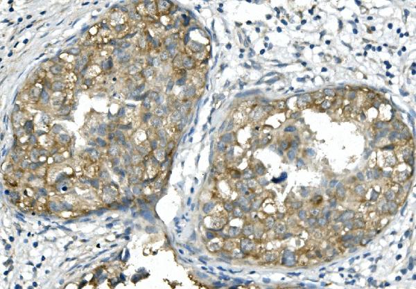
| Description | Anti-BubR1/BUB1B Antibody Picoband™ (monoclonal, 5I7) . Tested in Flow Cytometry, IF, IHC, ICC, WB applications. This antibody reacts with Human. |
| Reconstitution | Add 0.2ml of distilled water will yield a concentration of 500ug/ml. |
| Gene ID | 701 |
|---|---|
| Other Names | Mitotic checkpoint serine/threonine-protein kinase BUB1 beta, 2.7.11.1, MAD3/BUB1-related protein kinase, hBUBR1, Mitotic checkpoint kinase MAD3L, Protein SSK1, BUB1B, BUBR1, MAD3L, SSK1 |
| Calculated MW | 130 kDa |
| Application Details | Western blot, 0.1-0.5 µg/ml, Human Immunohistochemistry (Paraffin-embedded Section), 0.5-1 µg/ml, Human Immunocytochemistry/Immunofluorescence, 2 µg/ml, Human Flow Cytometry, 1-3 µg/1x10^6 cells, Human |
| Subcellular Localization | Centrosome. Nucleus. Cytoplasm. Kinetochore. |
| Tissue Specificity | Highly expressed in thymus followed by spleen. Preferentially expressed in tissues with a high mitotic index. |
| Contents | Each vial contains 4mg Trehalose, 0.9mg NaCl, 0.2mg Na2HPO4, 0.05mg NaN3. |
| Clone Names | Clone: 5I7 |
| Immunogen | E.coli-derived human BubR1/BUB1B recombinant protein (Position: K26-E448). |
| Purification | Immunogen affinity purified. |
| Cross Reactivity | No cross-reactivity with other proteins. |
| Storage | Store at -20˚C for one year from date of receipt. After reconstitution, at 4˚C for one month. It can also be aliquotted and stored frozen at -20˚C for six months. Avoid repeated freeze-thaw cycles. |
| Name | BUB1B |
|---|---|
| Synonyms | BUBR1, MAD3L, SSK1 |
| Function | Essential component of the mitotic checkpoint. Required for normal mitosis progression. The mitotic checkpoint delays anaphase until all chromosomes are properly attached to the mitotic spindle. One of its checkpoint functions is to inhibit the activity of the anaphase- promoting complex/cyclosome (APC/C) by blocking the binding of CDC20 to APC/C, independently of its kinase activity. The other is to monitor kinetochore activities that depend on the kinetochore motor CENPE. Required for kinetochore localization of CENPE. Negatively regulates PLK1 activity in interphase cells and suppresses centrosome amplification. Also implicated in triggering apoptosis in polyploid cells that exit aberrantly from mitotic arrest. May play a role for tumor suppression. |
| Cellular Location | Cytoplasm. Nucleus. Chromosome, centromere, kinetochore. Cytoplasm, cytoskeleton, microtubule organizing center, centrosome. Note=Cytoplasmic in interphase cells. Associates with the kinetochores in early prophase. Kinetochore localization requires BUB1, PLK1 and KNL1 |
| Tissue Location | Highly expressed in thymus followed by spleen. Preferentially expressed in tissues with a high mitotic index |

Thousands of laboratories across the world have published research that depended on the performance of antibodies from Abcepta to advance their research. Check out links to articles that cite our products in major peer-reviewed journals, organized by research category.
info@abcepta.com, and receive a free "I Love Antibodies" mug.

Provided below are standard protocols that you may find useful for product applications.
Background
Mitotic checkpoint serine/threonine-protein kinase BUB1 betais anenzymethat in humans is encoded by theBUB1Bgene. This gene encodes a kinase involved in spindle checkpoint function. The protein has been localized to the kinetochore and plays a role in the inhibition of the anaphase-promoting complex/cyclosome (APC/C), delaying the onset of anaphase and ensuring proper chromosome segregation. Impaired spindle checkpoint function has been found in many forms of cancer.
If you have used an Abcepta product and would like to share how it has performed, please click on the "Submit Review" button and provide the requested information. Our staff will examine and post your review and contact you if needed.
If you have any additional inquiries please email technical services at tech@abcepta.com.
Ordering Information
Other Products
Shipping Information
