


 Foundational characteristics of cancer include proliferation, angiogenesis, migration, evasion of apoptosis, and cellular immortality. Find key markers for these cellular processes and antibodies to detect them.
 The SUMOplot™ Analysis Program predicts and scores sumoylation sites in your protein. SUMOylation is a post-translational modification involved in various cellular processes, such as nuclear-cytosolic transport, transcriptional regulation, apoptosis, protein stability, response to stress, and progression through the cell cycle.
 The Autophagy Receptor Motif Plotter predicts and scores autophagy receptor binding sites in your protein. Identifying proteins connected to this pathway is critical to understanding the role of autophagy in physiological as well as pathological processes such as development, differentiation, neurodegenerative diseases, stress, infection, and cancer.
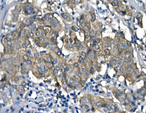
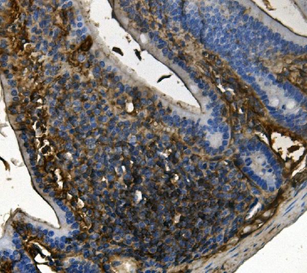
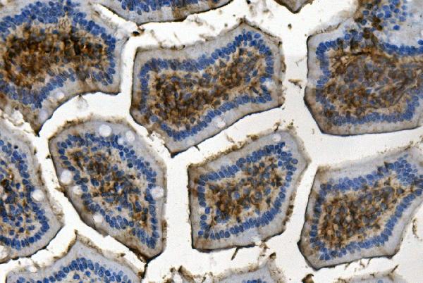
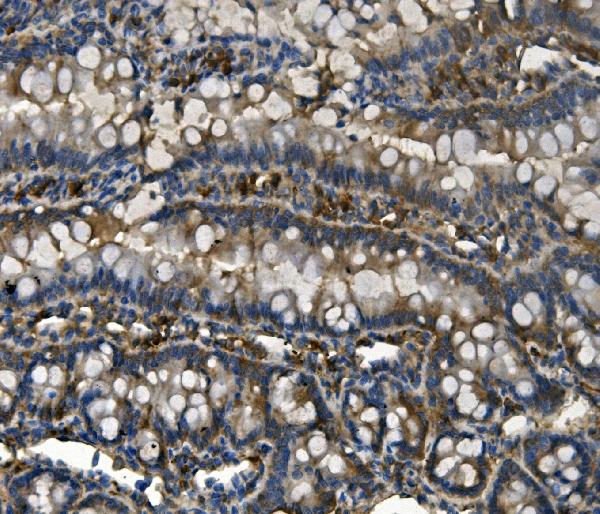
Anti-RENT1/hUPF1 Antibody Picoband™ (monoclonal, 11E7)

- SPECIFICATION
- CITATIONS
- PROTOCOLS
- BACKGROUND

Application
| WB, IHC, IF, ICC |
|---|---|
| Primary Accession | Q92900 |
| Host | Mouse |
| Isotype | Mouse IgG2b |
| Reactivity | Rat, Human, Mouse |
| Clonality | Monoclonal |
| Format | Lyophilized |
| Description | Anti-RENT1/hUPF1 Antibody Picoband™ (monoclonal, 11E7) . Tested in IF, IHC, ICC, WB applications. This antibody reacts with Human, Mouse, Rat. |
| Reconstitution | Add 0.2ml of distilled water will yield a concentration of 500ug/ml. |
| Gene ID | 5976 |
|---|---|
| Other Names | Regulator of nonsense transcripts 1 {ECO:0000312|HGNC:HGNC:9962}, 3.6.4.12, 3.6.4.13, ATP-dependent helicase RENT1, Up-frameshift suppressor 1 homolog, hUpf1, UPF1 (HGNC:9962) |
| Calculated MW | 130 kDa |
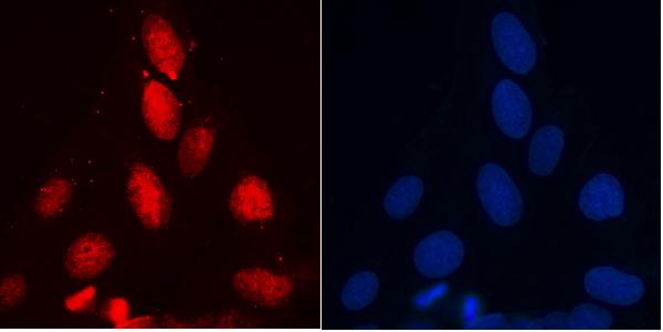
| Application Details | Western blot, 0.1-0.5 µg/ml, Human, Mouse, Rat Immunohistochemistry (Paraffin-embedded Section), 0.5-1 µg/ml, Human, Mouse, Rat Immunocytochemistry/Immunofluorescence, 2 µg/ml, Human |
| Subcellular Localization | Nucleus. Cytoplasm. P-body. |
| Tissue Specificity | Ubiquitous. |
| Contents | Each vial contains 4mg Trehalose, 0.9mg NaCl, 0.2mg Na2HPO4, 0.05mg NaN3. |
| Clone Names | Clone: 11E7 |
| Immunogen | A synthetic peptide corresponding to a sequence in the middle region of human RENT1/hUPF1, identical to the related mouse and rat sequences. |
| Purification | Immunogen affinity purified. |
| Cross Reactivity | No cross-reactivity with other proteins. |
| Storage | Store at -20˚C for one year from date of receipt. After reconstitution, at 4˚C for one month. It can also be aliquotted and stored frozen at -20˚C for six months. Avoid repeated freeze-thaw cycles. |
| Name | UPF1 (HGNC:9962) |
|---|---|
| Function | RNA-dependent helicase required for nonsense-mediated decay (NMD) of aberrant mRNAs containing premature stop codons and modulates the expression level of normal mRNAs (PubMed:11163187, PubMed:16086026, PubMed:18172165, PubMed:21145460, PubMed:21419344, PubMed:24726324). Is recruited to mRNAs upon translation termination and undergoes a cycle of phosphorylation and dephosphorylation; its phosphorylation appears to be a key step in NMD (PubMed:11544179, PubMed:25220460). Recruited by release factors to stalled ribosomes together with the SMG1C protein kinase complex to form the transient SURF (SMG1-UPF1-eRF1-eRF3) complex (PubMed:19417104). In EJC-dependent NMD, the SURF complex associates with the exon junction complex (EJC) (located 50-55 or more nucleotides downstream from the termination codon) through UPF2 and allows the formation of an UPF1-UPF2-UPF3 surveillance complex which is believed to activate NMD (PubMed:21419344). Phosphorylated UPF1 is recognized by EST1B/SMG5, SMG6 and SMG7 which are thought to provide a link to the mRNA degradation machinery involving exonucleolytic and endonucleolytic pathways, and to serve as adapters to protein phosphatase 2A (PP2A), thereby triggering UPF1 dephosphorylation and allowing the recycling of NMD factors (PubMed:12554878). UPF1 can also activate NMD without UPF2 or UPF3, and in the absence of the NMD-enhancing downstream EJC indicative for alternative NMD pathways (PubMed:18447585). Plays a role in replication-dependent histone mRNA degradation at the end of phase S; the function is independent of UPF2 (PubMed:16086026, PubMed:18172165). For the recognition of premature termination codons (PTC) and initiation of NMD a competitive interaction between UPF1 and PABPC1 with the ribosome-bound release factors is proposed (PubMed:18447585, PubMed:25220460). The ATPase activity of UPF1 is required for disassembly of mRNPs undergoing NMD (PubMed:21145460). Together with UPF2 and dependent on TDRD6, mediates the degradation of mRNA harboring long 3'UTR by inducing the NMD machinery (By similarity). Also capable of unwinding double-stranded DNA and translocating on single-stranded DNA (PubMed:30218034). |
| Cellular Location | Cytoplasm. Cytoplasm, P-body. Nucleus. Cytoplasm, perinuclear region {ECO:0000250|UniProtKB:Q9EPU0}. Note=Hyperphosphorylated form is targeted to the P-body, while unphosphorylated protein is distributed throughout the cytoplasm. Localized in the chromatoid bodies of round spermatids (By similarity). {ECO:0000250|UniProtKB:Q9EPU0} |
| Tissue Location | Ubiquitous. |

Thousands of laboratories across the world have published research that depended on the performance of antibodies from Abcepta to advance their research. Check out links to articles that cite our products in major peer-reviewed journals, organized by research category.
info@abcepta.com, and receive a free "I Love Antibodies" mug.

Provided below are standard protocols that you may find useful for product applications.
Background
Regulator of nonsense transcripts 1 is a protein that in humans is encoded by the UPF1 gene. This gene encodes a protein that is part of a post-splicing multiprotein complex involved in both mRNA nuclear export and mRNA surveillance. mRNA surveillance detects exported mRNAs with truncated open reading frames and initiates nonsense-mediated mRNA decay (NMD). When translation ends upstream from the last exon-exon junction, this triggers NMD to degrade mRNAs containing premature stop codons. And this protein is located only in the cytoplasm. When translation ends, it interacts with the protein that is a functional homolog of yeast Upf2p to trigger mRNA decapping. Use of multiple polyadenylation sites has been noted for this gene. Alternative splicing results in multiple transcript variants.
If you have used an Abcepta product and would like to share how it has performed, please click on the "Submit Review" button and provide the requested information. Our staff will examine and post your review and contact you if needed.
If you have any additional inquiries please email technical services at tech@abcepta.com.
Ordering Information
Other Products
Shipping Information
