


 Foundational characteristics of cancer include proliferation, angiogenesis, migration, evasion of apoptosis, and cellular immortality. Find key markers for these cellular processes and antibodies to detect them.
 The SUMOplot™ Analysis Program predicts and scores sumoylation sites in your protein. SUMOylation is a post-translational modification involved in various cellular processes, such as nuclear-cytosolic transport, transcriptional regulation, apoptosis, protein stability, response to stress, and progression through the cell cycle.
 The Autophagy Receptor Motif Plotter predicts and scores autophagy receptor binding sites in your protein. Identifying proteins connected to this pathway is critical to understanding the role of autophagy in physiological as well as pathological processes such as development, differentiation, neurodegenerative diseases, stress, infection, and cancer.
Anti-DDX5 Antibody Picoband™ (monoclonal, 3F9)

- SPECIFICATION
- CITATIONS
- PROTOCOLS
- BACKGROUND

Application
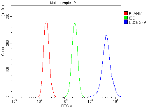
| WB, IHC, IHC-F, IF, ICC, FC |
|---|---|
| Primary Accession | P17844 |
| Host | Mouse |
| Isotype | Mouse IgG2b |
| Reactivity | Rat, Human, Mouse |
| Clonality | Monoclonal |
| Format | Lyophilized |
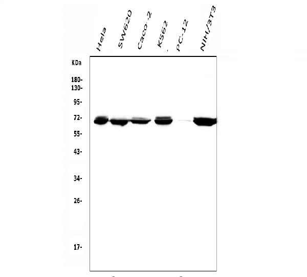
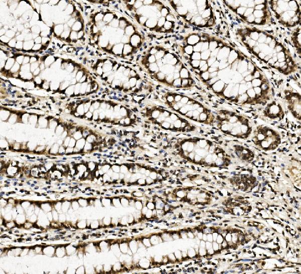
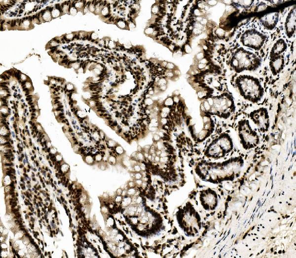
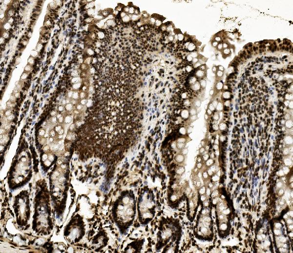
| Description | Anti-DDX5 Antibody Picoband™ (monoclonal, 3F9) . Tested in Flow Cytometry, IF, IHC, IHC-F, ICC, WB applications. This antibody reacts with Human, Mouse, Rat. |
| Reconstitution | Add 0.2ml of distilled water will yield a concentration of 500ug/ml. |
| Gene ID | 1655 |
|---|---|
| Other Names | Probable ATP-dependent RNA helicase DDX5, 3.6.4.13, DEAD box protein 5, RNA helicase p68, DDX5, G17P1, HELR, HLR1 |
| Calculated MW | 71 kDa |
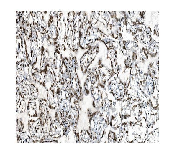
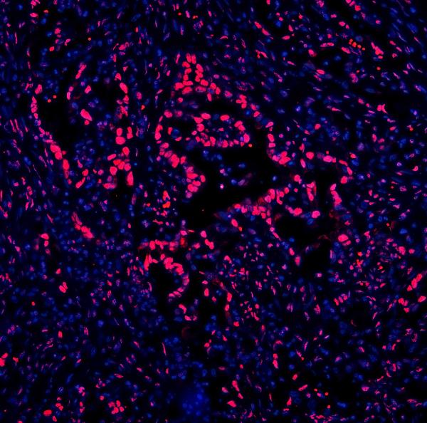
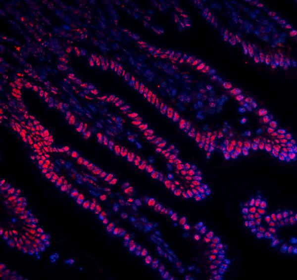
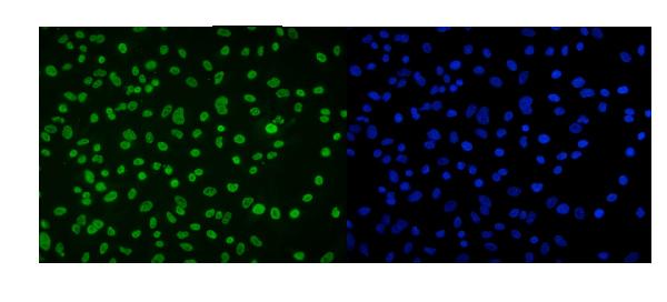
| Application Details | Western blot, 0.1-0.5 µg/ml, Human, Mouse, Rat Immunohistochemistry (Paraffin-embedded Section), 0.5-1 µg/ml, Human, Mouse, Rat Immunohistochemistry (Frozen Section), 0.5-1 µg/ml, Human Immunocytochemistry/Immunofluorescence, 2 µg/ml, Human Immunofluorescence, 2 µg/ml, Human, Mouse Flow Cytometry, 1-3 µg/1x10^6 cells, Human |
| Subcellular Localization | Nucleus. Spliceosome. |
| Contents | Each vial contains 4mg Trehalose, 0.9mg NaCl, 0.2mg Na2HPO4, 0.05mg NaN3. |
| Clone Names | Clone: 3F9 |
| Immunogen | E.coli-derived human DDX5 recombinant protein (Position: R85-K328). |
| Purification | Immunogen affinity purified. |
| Cross Reactivity | No cross-reactivity with other proteins. |
| Storage | Store at -20˚C for one year from date of receipt. After reconstitution, at 4˚C for one month. It can also be aliquotted and stored frozen at -20˚C for six months. Avoid repeated freeze-thaw cycles. |
| Name | DDX5 |
|---|---|
| Synonyms | G17P1, HELR, HLR1 |
| Function | Involved in the alternative regulation of pre-mRNA splicing; its RNA helicase activity is necessary for increasing tau exon 10 inclusion and occurs in a RBM4-dependent manner. Binds to the tau pre- mRNA in the stem-loop region downstream of exon 10. The rate of ATP hydrolysis is highly stimulated by single-stranded RNA. Involved in transcriptional regulation; the function is independent of the RNA helicase activity. Transcriptional coactivator for androgen receptor AR but probably not ESR1. Synergizes with DDX17 and SRA1 RNA to activate MYOD1 transcriptional activity and involved in skeletal muscle differentiation. Transcriptional coactivator for p53/TP53 and involved in p53/TP53 transcriptional response to DNA damage and p53/TP53- dependent apoptosis. Transcriptional coactivator for RUNX2 and involved in regulation of osteoblast differentiation. Acts as a transcriptional repressor in a promoter-specific manner; the function probably involves association with histone deacetylases, such as HDAC1. As component of a large PER complex is involved in the inhibition of 3' transcriptional termination of circadian target genes such as PER1 and NR1D1 and the control of the circadian rhythms. |
| Cellular Location | Nucleus. Nucleus, nucleolus Nucleus speckle. Cytoplasm. Note=During the G0 phase, predominantly located in the nucleus. Cytoplasmic levels increase during the G1/S phase. During the M phase, located at the vicinity of the condensed chromosomes. At G1, localizes in the cytoplasm |

Thousands of laboratories across the world have published research that depended on the performance of antibodies from Abcepta to advance their research. Check out links to articles that cite our products in major peer-reviewed journals, organized by research category.
info@abcepta.com, and receive a free "I Love Antibodies" mug.

Provided below are standard protocols that you may find useful for product applications.
Background
DDX5 (DEAD/H BOX 5), also known as HLR1 or G17P1, is an enzyme that in humans is encoded by the DDX5 gene. The p68 protein is a proliferation-associated nuclear antigen first identified through its highly specific cross-reaction with the simian virus 40 tumor antigen (Iggo et al., 1989). Subsequently, homology to eukaryotic translation initiation factor was found, and amino acid sequence blocks characteristic of a large superfamily of proteins with putative helicase activity were demonstrated. Brody et al. (1995) confirmed that this gene is located on chromosome 17 in the region of the BRCA1 gene at 17q21. By immunoprecipitation analysis, Caretti et al. (2006) found that p68, p72 (DDX17), and the noncoding RNA SRA (SRA1) associated with MYOD (MYOD1) in MYOD-transfected HeLa cells.
If you have used an Abcepta product and would like to share how it has performed, please click on the "Submit Review" button and provide the requested information. Our staff will examine and post your review and contact you if needed.
If you have any additional inquiries please email technical services at tech@abcepta.com.
Ordering Information
Other Products
Shipping Information
