


 Foundational characteristics of cancer include proliferation, angiogenesis, migration, evasion of apoptosis, and cellular immortality. Find key markers for these cellular processes and antibodies to detect them.
 The SUMOplot™ Analysis Program predicts and scores sumoylation sites in your protein. SUMOylation is a post-translational modification involved in various cellular processes, such as nuclear-cytosolic transport, transcriptional regulation, apoptosis, protein stability, response to stress, and progression through the cell cycle.
 The Autophagy Receptor Motif Plotter predicts and scores autophagy receptor binding sites in your protein. Identifying proteins connected to this pathway is critical to understanding the role of autophagy in physiological as well as pathological processes such as development, differentiation, neurodegenerative diseases, stress, infection, and cancer.
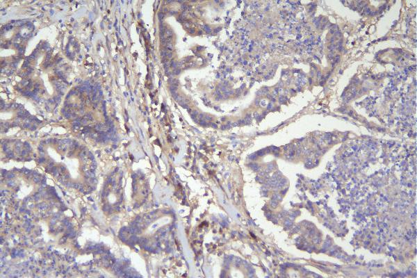
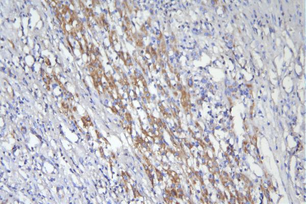
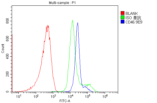
Anti-CD46 Antibody Picoband™ (monoclonal, 9E9)

- SPECIFICATION
- CITATIONS
- PROTOCOLS
- BACKGROUND

Application
| WB, IHC, FC |
|---|---|
| Primary Accession | P15529 |
| Host | Mouse |
| Isotype | Mouse IgG1 |
| Reactivity | Human |
| Clonality | Monoclonal |
| Format | Lyophilized |
| Description | Anti-CD46 Antibody Picoband™ (monoclonal, 9E9) . Tested in Flow Cytometry, IHC, WB applications. This antibody reacts with Human. |
| Reconstitution | Add 0.2ml of distilled water will yield a concentration of 500 µg/ml. |
| Gene ID | 4179 |
|---|---|
| Other Names | Membrane cofactor protein, TLX, Trophoblast leukocyte common antigen, CD46, CD46, MCP, MIC10 |
| Calculated MW | 50-70 kDa |
| Application Details | Western blot, 0.1-0.5 µg/ml, Human Immunohistochemistry (Paraffin-embedded Section), 0.5-1 µg/ml, Human Flow Cytometry, 1-3 µg/1x10^6 cells, Human |
| Tissue Specificity | Expressed by all cells except erythrocytes. |
| Protein Name | Membrane cofactor protein |
| Contents | Each vial contains 4mg Trehalose, 0.9mg NaCl, 0.2mg Na2HPO4, 0.05mg NaN3. |
| Clone Names | Clone: 9E9 |
| Immunogen | A synthetic peptide corresponding to a sequence at the C-terminus of human CD46. |
| Purification | Immunogen affinity purified. |
| Cross Reactivity | No cross-reactivity with other proteins. |
| Storage | Store at -20˚C for one year from date of receipt. After reconstitution, at 4˚C for one month. It can also be aliquotted and stored frozen at -20˚C for six months. Avoid repeated freeze-thaw cycles. |
| Name | CD46 |
|---|---|
| Synonyms | MCP, MIC10 |
| Function | Acts as a cofactor for complement factor I, a serine protease which protects autologous cells against complement-mediated injury by cleaving C3b and C4b deposited on host tissue. May be involved in the fusion of the spermatozoa with the oocyte during fertilization. Also acts as a costimulatory factor for T-cells which induces the differentiation of CD4+ into T-regulatory 1 cells. T-regulatory 1 cells suppress immune responses by secreting interleukin-10, and therefore are thought to prevent autoimmunity. |
| Cellular Location | Cytoplasmic vesicle, secretory vesicle, acrosome inner membrane; Single-pass type I membrane protein. Note=Inner acrosomal membrane of spermatozoa. Internalized upon binding of Measles virus, Herpesvirus 6 or Neisseria gonorrhoeae, which results in an increased susceptibility of infected cells to complement-mediated injury. In cancer cells or cells infected by Neisseria, shedding leads to a soluble peptide |
| Tissue Location | Expressed by all cells except erythrocytes. |

Thousands of laboratories across the world have published research that depended on the performance of antibodies from Abcepta to advance their research. Check out links to articles that cite our products in major peer-reviewed journals, organized by research category.
info@abcepta.com, and receive a free "I Love Antibodies" mug.

Provided below are standard protocols that you may find useful for product applications.
Background
CD46 complement regulatory protein also known as CD46 (cluster of differentiation 46) and Membrane Cofactor Protein is a protein which in humans is encoded by the CD46 gene. The protein encoded by this gene is a type I membrane protein and is a regulatory part of the complement system. And the encoded protein has cofactor activity for inactivation of complement components C3b and C4b by serum factor I, which protects the host cell from damage by complement. In addition, the encoded protein can act as a receptor for the Edmonston strain of measles virus, human herpesvirus-6, and type IV pili of pathogenic Neisseria. Finally, the protein encoded by this gene may be involved in the fusion of the spermatozoa with the oocyte during fertilization. Mutations at this locus have been associated with susceptibility to hemolytic uremic syndrome. Alternatively spliced transcript variants encoding different isoforms have been described.
If you have used an Abcepta product and would like to share how it has performed, please click on the "Submit Review" button and provide the requested information. Our staff will examine and post your review and contact you if needed.
If you have any additional inquiries please email technical services at tech@abcepta.com.
Ordering Information
Other Products
Shipping Information
