


 Foundational characteristics of cancer include proliferation, angiogenesis, migration, evasion of apoptosis, and cellular immortality. Find key markers for these cellular processes and antibodies to detect them.
 The SUMOplot™ Analysis Program predicts and scores sumoylation sites in your protein. SUMOylation is a post-translational modification involved in various cellular processes, such as nuclear-cytosolic transport, transcriptional regulation, apoptosis, protein stability, response to stress, and progression through the cell cycle.
 The Autophagy Receptor Motif Plotter predicts and scores autophagy receptor binding sites in your protein. Identifying proteins connected to this pathway is critical to understanding the role of autophagy in physiological as well as pathological processes such as development, differentiation, neurodegenerative diseases, stress, infection, and cancer.
Anti-PRDX6 Antibody Picoband™ (monoclonal, 6I8)
- SPECIFICATION
- CITATIONS
- PROTOCOLS
- BACKGROUND

Application
| WB, IHC, IF, ICC, FC |
|---|---|
| Primary Accession | P30041 |
| Host | Mouse |
| Isotype | Mouse IgG2b |
| Reactivity | Rat, Human, Mouse |
| Clonality | Monoclonal |
| Format | Lyophilized |
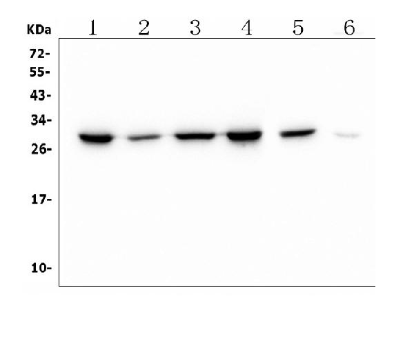
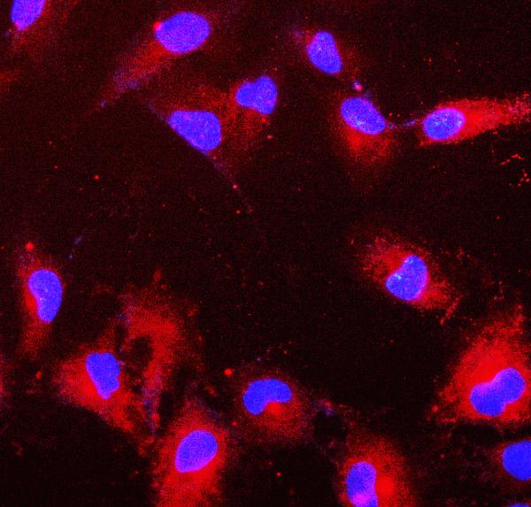
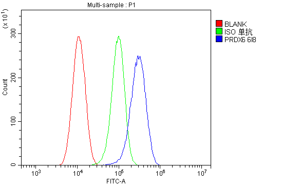
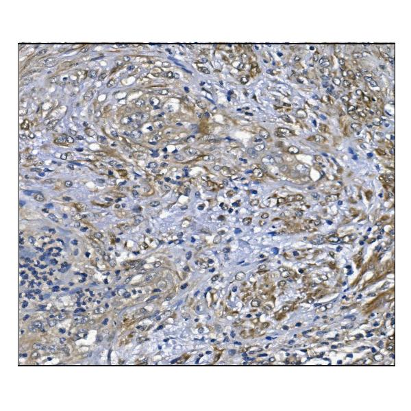
| Description | Anti-PRDX6 Antibody Picoband™ (monoclonal, 6I8) . Tested in Flow Cytometry, IF, IHC, ICC, WB applications. This antibody reacts with Human, Mouse, Rat. |
| Reconstitution | Add 0.2ml of distilled water will yield a concentration of 500 µg/ml. |
| Gene ID | 9588 |
|---|---|
| Other Names | Peroxiredoxin-6, 1.11.1.27, 1-Cys peroxiredoxin, 1-Cys PRX, 24 kDa protein, Acidic calcium-independent phospholipase A2, aiPLA2, 3.1.1.4, Antioxidant protein 2, Glutathione-dependent peroxiredoxin, Liver 2D page spot 40, Lysophosphatidylcholine acyltransferase 5, LPC acyltransferase 5, LPCAT-5, Lyso-PC acyltransferase 5, 2.3.1.23, Non-selenium glutathione peroxidase, NSGPx, Red blood cells page spot 12, PRDX6, AOP2, KIAA0106 |
| Calculated MW | 25035 MW KDa |
| Application Details | Western blot, 0.25-0.5 µg/ml, Human, Rat Immunohistochemistry (Paraffin-embedded Section), 0.5-1 µg/ml, Human Immunocytochemistry/Immunofluorescence, 2 µg/ml, Human Flow Cytometry, 1-3 µg/1x10^6 cells, Human |
| Subcellular Localization | Cytoplasm. Lysosome. Cytoplasmic vesicle. Also found in lung secretory organelles. |
| Protein Name | Peroxiredoxin-6 |
| Contents | Each vial contains 4mg Trehalose, 0.9mg NaCl, 0.2mg Na2HPO4, 0.05mg NaN3. |
| Clone Names | Clone: 6I8 |
| Immunogen | E.coli-derived human Peroxiredoxin 6 recombinant protein (Position: E15-P224). Human Peroxiredoxin 6 shares 90% and 91% amino acid (aa) sequence identity with mouse and rat Peroxiredoxin 6, respectively. |
| Purification | Immunogen affinity purified. |
| Cross Reactivity | No cross-reactivity with other proteins. |
| Storage | Store at -20˚C for one year from date of receipt. After reconstitution, at 4˚C for one month. It can also be aliquotted and stored frozen at -20˚C for six months. Avoid repeated freeze-thaw cycles. |
| Name | PRDX6 |
|---|---|
| Synonyms | AOP2, KIAA0106 |
| Function | Thiol-specific peroxidase that catalyzes the reduction of hydrogen peroxide and organic hydroperoxides to water and alcohols, respectively (PubMed:10893423, PubMed:9497358). Can reduce H(2)O(2) and short chain organic, fatty acid, and phospholipid hydroperoxides (PubMed:10893423). Also has phospholipase activity, can therefore either reduce the oxidized sn-2 fatty acyl group of phospholipids (peroxidase activity) or hydrolyze the sn-2 ester bond of phospholipids (phospholipase activity) (PubMed:10893423, PubMed:26830860). These activities are dependent on binding to phospholipids at acidic pH and to oxidized phospholipds at cytosolic pH (PubMed:10893423). Plays a role in cell protection against oxidative stress by detoxifying peroxides and in phospholipid homeostasis (PubMed:10893423). Exhibits acyl-CoA-dependent lysophospholipid acyltransferase which mediates the conversion of lysophosphatidylcholine (1-acyl-sn-glycero-3- phosphocholine or LPC) into phosphatidylcholine (1,2-diacyl-sn-glycero- 3-phosphocholine or PC) (PubMed:26830860). Shows a clear preference for LPC as the lysophospholipid and for palmitoyl CoA as the fatty acyl substrate (PubMed:26830860). |
| Cellular Location | Cytoplasm. Lysosome {ECO:0000250|UniProtKB:O35244}. Note=Also found in lung secretory organelles (lamellar bodies). {ECO:0000250|UniProtKB:O35244} |

Thousands of laboratories across the world have published research that depended on the performance of antibodies from Abcepta to advance their research. Check out links to articles that cite our products in major peer-reviewed journals, organized by research category.
info@abcepta.com, and receive a free "I Love Antibodies" mug.

Provided below are standard protocols that you may find useful for product applications.
Background
PRDX6 is also known as PRX, p29 or AOP2. The protein encoded by this gene is a member of the thiol-specific antioxidant protein family. This protein is a bifunctional enzyme with two distinct active sites. It is involved in redox regulation of the cell; it can reduce H (2)O (2) and short chain organic, fatty acid, and phospholipid hydroperoxides. It may play a role in the regulation of phospholipid turnover as well as in protection against oxidative injury.
If you have used an Abcepta product and would like to share how it has performed, please click on the "Submit Review" button and provide the requested information. Our staff will examine and post your review and contact you if needed.
If you have any additional inquiries please email technical services at tech@abcepta.com.
Ordering Information
Other Products
Shipping Information
