


 Foundational characteristics of cancer include proliferation, angiogenesis, migration, evasion of apoptosis, and cellular immortality. Find key markers for these cellular processes and antibodies to detect them.
 The SUMOplot™ Analysis Program predicts and scores sumoylation sites in your protein. SUMOylation is a post-translational modification involved in various cellular processes, such as nuclear-cytosolic transport, transcriptional regulation, apoptosis, protein stability, response to stress, and progression through the cell cycle.
 The Autophagy Receptor Motif Plotter predicts and scores autophagy receptor binding sites in your protein. Identifying proteins connected to this pathway is critical to understanding the role of autophagy in physiological as well as pathological processes such as development, differentiation, neurodegenerative diseases, stress, infection, and cancer.
Anti-Mitofusin 1 MFN1 Antibody Picoband™ (monoclonal, 3H3)
- SPECIFICATION
- CITATIONS
- PROTOCOLS
- BACKGROUND

Application
| WB, IF, ICC |
|---|---|
| Primary Accession | Q8IWA4 |
| Host | Mouse |
| Isotype | Mouse IgG2b |
| Reactivity | Human |
| Clonality | Monoclonal |
| Format | Lyophilized |
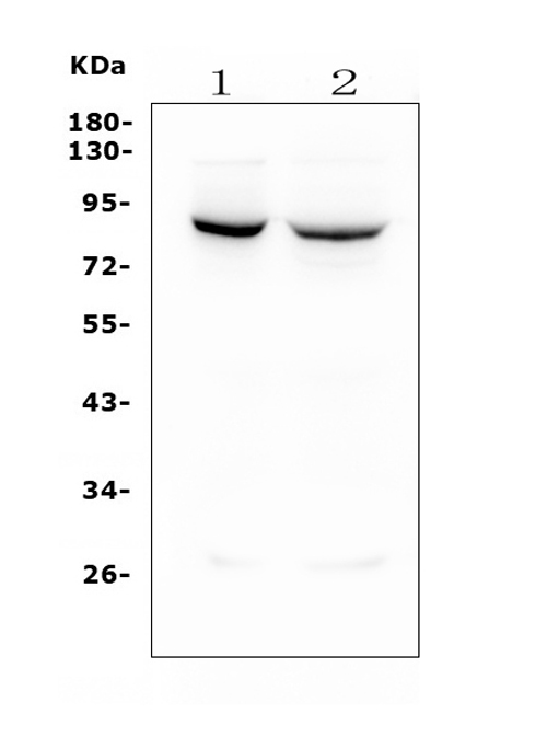
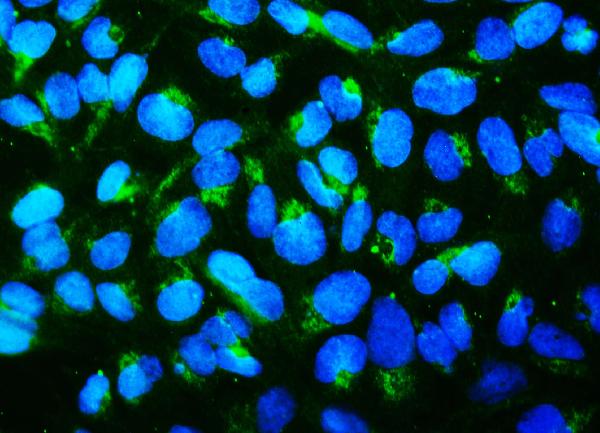
| Description | Anti-Mitofusin 1 MFN1 Antibody Picoband™ (monoclonal, 3H3) . Tested in IF, ICC, WB applications. This antibody reacts with Human. |
| Reconstitution | Add 0.2ml of distilled water will yield a concentration of 500 µg/ml. |
| Gene ID | 55669 |
|---|---|
| Other Names | Mitofusin-1, 3.6.5.-, Fzo homolog, Transmembrane GTPase MFN1, MFN1 |
| Calculated MW | 84 kDa |
| Application Details | Western blot, 0.1-0.5 µg/ml, Human Immunocytochemistry/Immunofluorescence, 2 µg/ml, Human |
| Subcellular Localization | Mitochondrion outer membrane ; Multi- pass membrane protein. |
| Tissue Specificity | Ubiquitous.Expressed at slightly higher level in kidney and heart.Isoform 2 may be overexpressed in some tumors,such as lung cancers. |
| Protein Name | Mitofusin-1 |
| Contents | Each vial contains 4mg Trehalose, 0.9mg NaCl, 0.2mg Na2HPO4, 0.05mg NaN3. |
| Clone Names | Clone: 3H3 |
| Immunogen | A synthetic peptide corresponding to a sequence at the N-terminal of human Mitofusin 1, different from the related mouse and rat sequences by one amino acid. |
| Purification | Immunogen affinity purified. |
| Cross Reactivity | No cross-reactivity with other proteins. |
| Storage | Store at -20˚C for one year from date of receipt. After reconstitution, at 4˚C for one month. It can also be aliquotted and stored frozen at -20˚C for six months. Avoid repeated freeze-thaw cycles. |
| Name | MFN1 |
|---|---|
| Function | Mitochondrial outer membrane GTPase that mediates mitochondrial clustering and fusion (PubMed:12475957, PubMed:12759376, PubMed:27920125, PubMed:28114303). Membrane clustering requires GTPase activity (PubMed:27920125). It may involve a major rearrangement of the coiled coil domains (PubMed:27920125, PubMed:28114303). Mitochondria are highly dynamic organelles, and their morphology is determined by the equilibrium between mitochondrial fusion and fission events (PubMed:12475957, PubMed:12759376). Overexpression induces the formation of mitochondrial networks (in vitro) (PubMed:12759376). Has low GTPase activity (PubMed:27920125, PubMed:28114303). |
| Cellular Location | Mitochondrion outer membrane; Multi-pass membrane protein |
| Tissue Location | Detected in kidney and heart (at protein level) (PubMed:12759376). Ubiquitous (PubMed:11950885, PubMed:12759376) Expressed at slightly higher level in kidney and heart (PubMed:12759376). Isoform 2 may be overexpressed in some tumors, such as lung cancers (PubMed:11751411). |

Thousands of laboratories across the world have published research that depended on the performance of antibodies from Abcepta to advance their research. Check out links to articles that cite our products in major peer-reviewed journals, organized by research category.
info@abcepta.com, and receive a free "I Love Antibodies" mug.

Provided below are standard protocols that you may find useful for product applications.
Background
Mitofusin-1 is a protein that in humans is encoded by the MFN1 gene. It is an 8090 kDa mitochondrial member of the dynamin family of molecules. It is ubiquitously expressed, and found in the outer mitochondrial membrane. The protein encoded by this gene is a mediator of mitochondrial fusion, and thereby contribute to the dynamic balance between fusion and fission that determines mitochondria morphology. MFN1 is known to form oligomers, either with itself or MFN2, and to undergo ubiquitination by MARCH5. MFN1 has two key domains. One is a coiledcoil region that mediates MFN1: MFN1/2 binding, and a second is a GTPase domain whose cleavage of GTP is necessary for membrane fusion. Overexpression of MFN1 caused perinuclear mitochondrial clustering.
If you have used an Abcepta product and would like to share how it has performed, please click on the "Submit Review" button and provide the requested information. Our staff will examine and post your review and contact you if needed.
If you have any additional inquiries please email technical services at tech@abcepta.com.
Ordering Information
Other Products
Shipping Information
