


 Foundational characteristics of cancer include proliferation, angiogenesis, migration, evasion of apoptosis, and cellular immortality. Find key markers for these cellular processes and antibodies to detect them.
 The SUMOplot™ Analysis Program predicts and scores sumoylation sites in your protein. SUMOylation is a post-translational modification involved in various cellular processes, such as nuclear-cytosolic transport, transcriptional regulation, apoptosis, protein stability, response to stress, and progression through the cell cycle.
 The Autophagy Receptor Motif Plotter predicts and scores autophagy receptor binding sites in your protein. Identifying proteins connected to this pathway is critical to understanding the role of autophagy in physiological as well as pathological processes such as development, differentiation, neurodegenerative diseases, stress, infection, and cancer.
Anti-HMG4 Antibody Picoband™ (monoclonal, 8H9)
- SPECIFICATION
- CITATIONS
- PROTOCOLS
- BACKGROUND

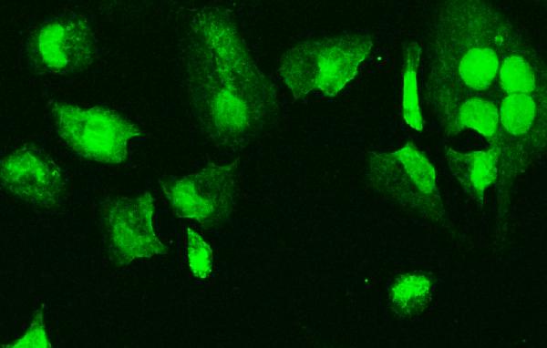
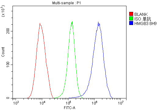
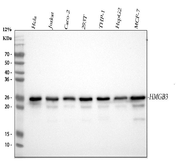
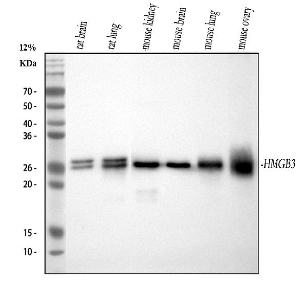
Application
| WB, IF, ICC, FC |
|---|---|
| Primary Accession | O15347 |
| Host | Mouse |
| Isotype | Mouse IgG2b |
| Reactivity | Rat, Human, Mouse |
| Clonality | Monoclonal |
| Format | Lyophilized |
| Description | Anti-HMG4 Antibody Picoband™ (monoclonal, 8H9) . Tested in Flow Cytometry, IF, ICC, WB applications. This antibody reacts with Human, Mouse, Rat. |
| Reconstitution | Add 0.2ml of distilled water will yield a concentration of 500 µg/ml. |
| Gene ID | 3149 |
|---|---|
| Other Names | High mobility group protein B3, High mobility group protein 2a, HMG-2a, High mobility group protein 4, HMG-4, HMGB3, HMG2A, HMG4 |
| Calculated MW | 23 kDa |
| Application Details | Western blot, 0.1-0.5 µg/ml, Human, Mouse, Rat Immunocytochemistry/Immunofluorescence, 2 µg/ml, Human Flow Cytometry, 1-3 µg/1x10^6 cells, Human |
| Subcellular Localization | Nucleus. Chromosome. Cytoplasm. |
| Tissue Specificity | Expressed predominantly in placenta. |
| Protein Name | High mobility group protein B3 |
| Contents | Each vial contains 4mg Trehalose, 0.9mg NaCl, 0.2mg Na2HPO4, 0.05mg NaN3. |
| Clone Names | Clone: 8H9 |
| Immunogen | A synthetic peptide corresponding to a sequence at the N-terminus of human HMG4, identical to the related mouse and rat sequences. |
| Purification | Immunogen affinity purified. |
| Cross Reactivity | No cross-reactivity with other proteins. |
| Storage | Store at -20˚C for one year from date of receipt. After reconstitution, at 4˚C for one month. It can also be aliquotted and stored frozen at -20˚C for six months. Avoid repeated freeze-thaw cycles. |
| Name | HMGB3 |
|---|---|
| Synonyms | HMG2A, HMG4 |
| Function | Multifunctional protein with various roles in different cellular compartments. May act in a redox sensitive manner. Associates with chromatin and binds DNA with a preference for non-canonical DNA structures such as single-stranded DNA. Can bend DNA and enhance DNA flexibility by looping thus providing a mechanism to promote activities on various gene promoters (By similarity). Proposed to be involved in the innate immune response to nucleic acids by acting as a cytoplasmic promiscuous immunogenic DNA/RNA sensor (By similarity). Negatively regulates B-cell and myeloid cell differentiation. In hematopoietic stem cells may regulate the balance between self-renewal and differentiation. Involved in negative regulation of canonical Wnt signaling (By similarity). |
| Cellular Location | Nucleus {ECO:0000250|UniProtKB:P40618, ECO:0000255|PROSITE-ProRule:PRU00267}. Chromosome Cytoplasm {ECO:0000250|UniProtKB:O54879} |
| Tissue Location | Expressed predominantly in placenta. |

Thousands of laboratories across the world have published research that depended on the performance of antibodies from Abcepta to advance their research. Check out links to articles that cite our products in major peer-reviewed journals, organized by research category.
info@abcepta.com, and receive a free "I Love Antibodies" mug.

Provided below are standard protocols that you may find useful for product applications.
Background
High-mobility group protein B, also known as HMG4, is a protein that in humans is encoded by the HMGB3 gene. This gene encodes a member of a family of proteins containing one or more high mobility group DNA-binding motifs. The encoded protein plays an important role in maintaining stem cell populations, and may be aberrantly expressed in tumor cells. A mutation in this gene was associated with microphthalmia, syndromic 13. There are numerous pseudogenes of this gene on multiple chromosomes. Alternative splicing results in multiple transcript variants.
If you have used an Abcepta product and would like to share how it has performed, please click on the "Submit Review" button and provide the requested information. Our staff will examine and post your review and contact you if needed.
If you have any additional inquiries please email technical services at tech@abcepta.com.
Ordering Information
Other Products
Shipping Information
