


 Foundational characteristics of cancer include proliferation, angiogenesis, migration, evasion of apoptosis, and cellular immortality. Find key markers for these cellular processes and antibodies to detect them.
 The SUMOplot™ Analysis Program predicts and scores sumoylation sites in your protein. SUMOylation is a post-translational modification involved in various cellular processes, such as nuclear-cytosolic transport, transcriptional regulation, apoptosis, protein stability, response to stress, and progression through the cell cycle.
 The Autophagy Receptor Motif Plotter predicts and scores autophagy receptor binding sites in your protein. Identifying proteins connected to this pathway is critical to understanding the role of autophagy in physiological as well as pathological processes such as development, differentiation, neurodegenerative diseases, stress, infection, and cancer.
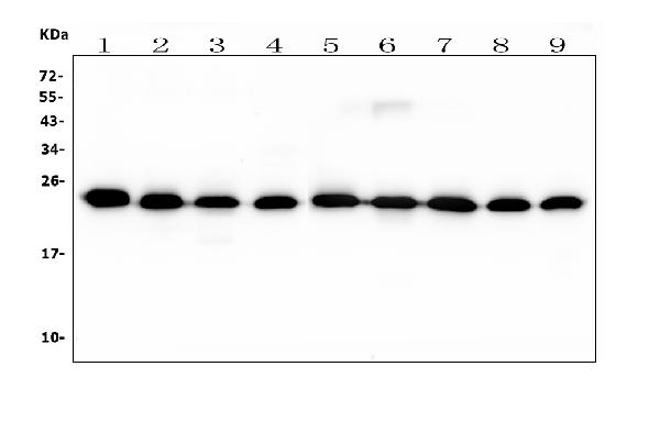
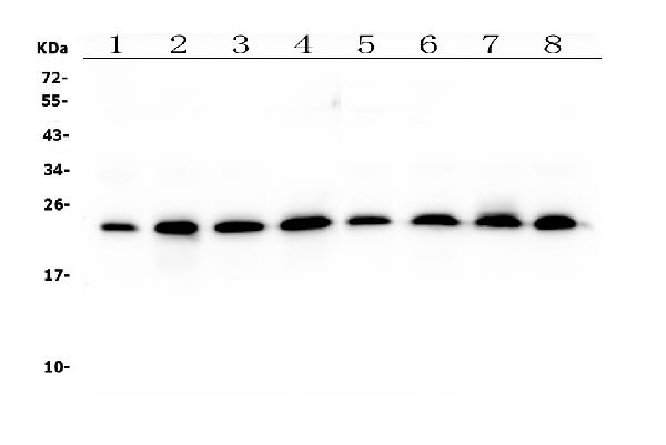
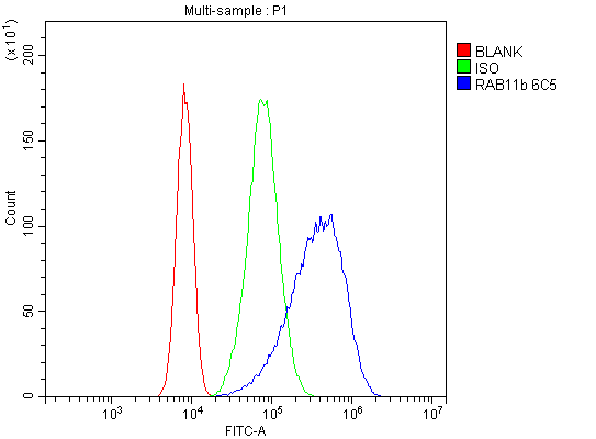
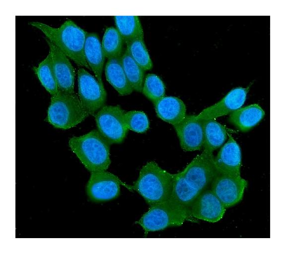
Anti-RAB11B Antibody Picoband™ (monoclonal, 6C5)
- SPECIFICATION
- CITATIONS
- PROTOCOLS
- BACKGROUND

Application
| WB, IF, ICC, FC |
|---|---|
| Primary Accession | Q15907 |
| Host | Mouse |
| Isotype | Mouse IgG2b |
| Reactivity | Rat, Human, Mouse |
| Clonality | Monoclonal |
| Format | Lyophilized |
| Description | Anti-RAB11B Antibody Picoband™ (monoclonal, 6C5) . Tested in Flow Cytometry, IF, ICC, WB applications. This antibody reacts with Human, Mouse, Rat. |
| Reconstitution | Add 0.2ml of distilled water will yield a concentration of 500 µg/ml. |
| Gene ID | 9230 |
|---|---|
| Other Names | Ras-related protein Rab-11B, 3.6.5.2, GTP-binding protein YPT3, RAB11B, YPT3 |
| Calculated MW | 24 kDa |
| Application Details | Western blot, 0.1-0.5 µg/ml, Human, Mouse, Rat Immunocytochemistry/Immunofluorescence, 2 µg/ml, Human Flow Cytometry, 1-3 µg/1x10^6 cells, Human |
| Subcellular Localization | Recycling endosome membrane;Lipid-anchor;Cytoplasmic side.Cytoplasmic vesicle,secretory vesicle,synaptic vesicle membrane;Lipid-anchor;Cytoplasmic side.Cytoplasmic vesicle,phagosome membrane;Lipid-anchor;Cytoplasmic side.Recruited to phagosomes containing S.aureus. |
| Protein Name | Ras-related protein Rab-11B |
| Contents | Each vial contains 4mg Trehalose, 0.9mg NaCl, 0.2mg Na2HPO4, 0.05mg NaN3. |
| Clone Names | Clone: 6C5 |
| Immunogen | A synthetic peptide corresponding to a sequence at the C-terminus of human RAB11B, which shares 97.4% and 100% amino acid (aa) sequence identity with mouse and rat RAB11B, respectively. |
| Purification | Immunogen affinity purified. |
| Cross Reactivity | No cross-reactivity with other proteins. |
| Storage | Store at -20˚C for one year from date of receipt. After reconstitution, at 4˚C for one month. It can also be aliquotted and stored frozen at -20˚C for six months. Avoid repeated freeze-thaw cycles. |
| Name | RAB11B |
|---|---|
| Synonyms | YPT3 |
| Function | The small GTPases Rab are key regulators of intracellular membrane trafficking, from the formation of transport vesicles to their fusion with membranes. Rabs cycle between an inactive GDP-bound form and an active GTP-bound form that is able to recruit to membranes different set of downstream effectors directly responsible for vesicle formation, movement, tethering and fusion. The small Rab GTPase RAB11B plays a role in endocytic recycling, regulating apical recycling of several transmembrane proteins including cystic fibrosis transmembrane conductance regulator/CFTR, epithelial sodium channel/ENaC, potassium voltage-gated channel, and voltage-dependent L-type calcium channel. May also regulate constitutive and regulated secretion, like insulin granule exocytosis. Required for melanosome transport and release from melanocytes. Also regulates V-ATPase intracellular transport in response to extracellular acidosis. Promotes Rabin8/RAB3IP preciliary vesicular trafficking to mother centriole by forming a ciliary targeting complex containing Rab11, ASAP1, Rabin8/RAB3IP, RAB11FIP3 and ARF4, thereby regulating ciliogenesis initiation (Probable). On the contrary, upon LPAR1 receptor signaling pathway activation, interaction with phosphorylated WDR44 prevents Rab11-RAB3IP-RAB11FIP3 complex formation and cilia growth (Probable). |
| Cellular Location | Recycling endosome membrane {ECO:0000250|UniProtKB:P46638}; Lipid-anchor {ECO:0000250|UniProtKB:P46638}; Cytoplasmic side {ECO:0000250|UniProtKB:P46638}. Cytoplasmic vesicle, secretory vesicle, synaptic vesicle membrane {ECO:0000250|UniProtKB:O35509}; Lipid-anchor {ECO:0000250|UniProtKB:O35509}; Cytoplasmic side {ECO:0000250|UniProtKB:O35509}. Cytoplasmic vesicle, phagosome membrane; Lipid-anchor; Cytoplasmic side. Note=Recruited to phagosomes containing S.aureus. |

Thousands of laboratories across the world have published research that depended on the performance of antibodies from Abcepta to advance their research. Check out links to articles that cite our products in major peer-reviewed journals, organized by research category.
info@abcepta.com, and receive a free "I Love Antibodies" mug.

Provided below are standard protocols that you may find useful for product applications.
Background
Ras-related protein Rab-11B is a protein that in humans is encoded by the RAB11B gene. It is mapped to 19p13.2. The Ras superfamily of small GTP-binding proteins, which includes the Ras, Ral, Rho, Rap, and Rab families, is involved in controlling a diverse set of essential cellular functions. The Rab family, including RAB11B, appears to play a critical role in regulating exocytotic and endocytotic pathways.
If you have used an Abcepta product and would like to share how it has performed, please click on the "Submit Review" button and provide the requested information. Our staff will examine and post your review and contact you if needed.
If you have any additional inquiries please email technical services at tech@abcepta.com.
Ordering Information
Other Products
Shipping Information
