


 Foundational characteristics of cancer include proliferation, angiogenesis, migration, evasion of apoptosis, and cellular immortality. Find key markers for these cellular processes and antibodies to detect them.
 The SUMOplot™ Analysis Program predicts and scores sumoylation sites in your protein. SUMOylation is a post-translational modification involved in various cellular processes, such as nuclear-cytosolic transport, transcriptional regulation, apoptosis, protein stability, response to stress, and progression through the cell cycle.
 The Autophagy Receptor Motif Plotter predicts and scores autophagy receptor binding sites in your protein. Identifying proteins connected to this pathway is critical to understanding the role of autophagy in physiological as well as pathological processes such as development, differentiation, neurodegenerative diseases, stress, infection, and cancer.
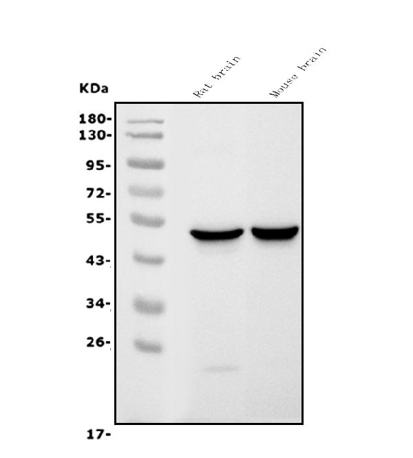
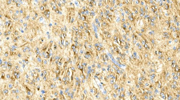
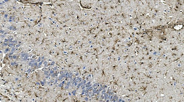
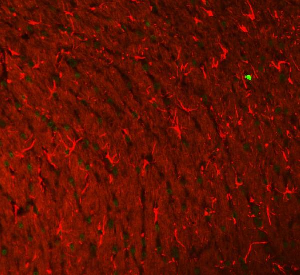
Anti-GFAP Antibody Picoband™ (monoclonal, 3F2)
- SPECIFICATION
- CITATIONS
- PROTOCOLS
- BACKGROUND

Application
| WB, IHC, IF |
|---|---|
| Primary Accession | P14136 |
| Host | Mouse |
| Isotype | Mouse IgG1 |
| Reactivity | Rat, Human, Mouse |
| Clonality | Monoclonal |
| Format | Lyophilized |
| Description | Anti-GFAP Antibody Picoband™ (monoclonal, 3F2) . Tested in IF, IHC, WB applications. This antibody reacts with Human, Mouse, Rat. |
| Reconstitution | Add 0.2ml of distilled water will yield a concentration of 500 µg/ml. |
| Gene ID | 2670 |
|---|---|
| Other Names | Glial fibrillary acidic protein, GFAP, GFAP |
| Calculated MW | 50 kDa |
| Application Details | Western blot, 0.1-0.5 µg/ml, Mouse, rat Immunohistochemistry (Paraffin-embedded Section), 0.5-1 µg/ml, Human, Rat Immunofluorescence, 5 µg/ml, Rat |
| Protein Name | Glial fibrillary acidic protein |
| Contents | Each vial contains 4mg Trehalose, 0.9mg NaCl, 0.2mg Na2HPO4, 0.05mg NaN3. |
| Clone Names | Clone: 3F2 |
| Immunogen | E.coli-derived human GFAP recombinant protein (Position: Q93-M432). Human GFAP shares 94% amino acid (aa) sequence identity with both mouse and rat GFAP. |
| Purification | Immunogen affinity purified. |
| Cross Reactivity | No cross-reactivity with other proteins. |
| Storage | Store at -20˚C for one year from date of receipt. After reconstitution, at 4˚C for one month. It can also be aliquotted and stored frozen at -20˚C for six months. Avoid repeated freeze-thaw cycles. |
| Name | GFAP |
|---|---|
| Function | GFAP, a class-III intermediate filament, is a cell-specific marker that, during the development of the central nervous system, distinguishes astrocytes from other glial cells. |
| Cellular Location | Cytoplasm. Note=Associated with intermediate filaments |
| Tissue Location | Expressed in cells lacking fibronectin. |

Thousands of laboratories across the world have published research that depended on the performance of antibodies from Abcepta to advance their research. Check out links to articles that cite our products in major peer-reviewed journals, organized by research category.
info@abcepta.com, and receive a free "I Love Antibodies" mug.

Provided below are standard protocols that you may find useful for product applications.
Background
Glial fibrillary acidic protein (GFAP) is a protein that is encoded by the GFAP gene in humans. It is an intermediate filament (IF) protein that is expressed by numerous cell types of the central nervous system (CNS) including astrocytes, and ependymal cells. It is mapped to 17q21.31. GFAP is closely related to its non-epithelial family members, vimentin, desmin, and peripherin, which are all involved in the structure and function of the cell’s cytoskeleton. GFAP is thought to help to maintain astrocyte mechanical strength, as well as the shape of cells. This gene has been shown to play a role in mitosis by adjusting the filament network present in the cell. GFAP is necessary for many critical roles in the CNS. What’s more, GFAP also plays a role in astrocyte-neuron interactions as well as cell-cell communication.
If you have used an Abcepta product and would like to share how it has performed, please click on the "Submit Review" button and provide the requested information. Our staff will examine and post your review and contact you if needed.
If you have any additional inquiries please email technical services at tech@abcepta.com.
Ordering Information
Other Products
Shipping Information
