


 Foundational characteristics of cancer include proliferation, angiogenesis, migration, evasion of apoptosis, and cellular immortality. Find key markers for these cellular processes and antibodies to detect them.
 The SUMOplot™ Analysis Program predicts and scores sumoylation sites in your protein. SUMOylation is a post-translational modification involved in various cellular processes, such as nuclear-cytosolic transport, transcriptional regulation, apoptosis, protein stability, response to stress, and progression through the cell cycle.
 The Autophagy Receptor Motif Plotter predicts and scores autophagy receptor binding sites in your protein. Identifying proteins connected to this pathway is critical to understanding the role of autophagy in physiological as well as pathological processes such as development, differentiation, neurodegenerative diseases, stress, infection, and cancer.
Anti-CD59 Antibody Picoband™(monoclonal, 3C10)
- SPECIFICATION
- CITATIONS
- PROTOCOLS
- BACKGROUND

Application
| WB, IHC, IF, ICC, FC |
|---|---|
| Primary Accession | P13987 |
| Host | Mouse |
| Isotype | Mouse IgG2b |
| Reactivity | Human |
| Clonality | Monoclonal |
| Format | Lyophilized |
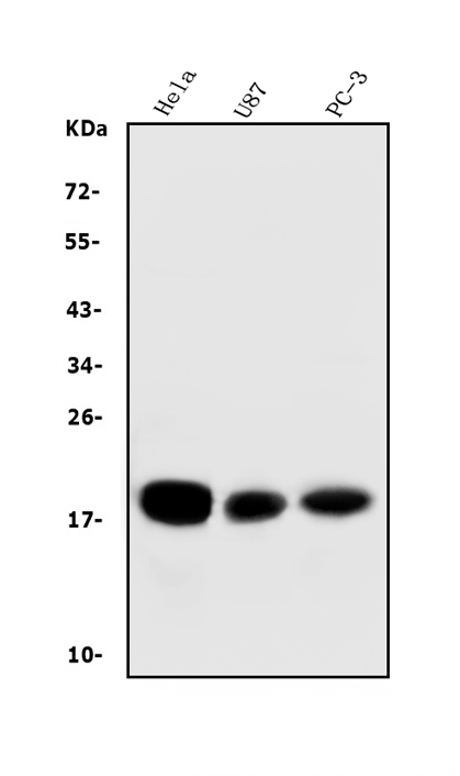
| Description | Anti-CD59 Antibody Picoband™ (monoclonal, 3C10) . Tested in Flow Cytometry, IF, IHC, ICC, WB applications. This antibody reacts with Human. |
| Reconstitution | Add 0.2ml of distilled water will yield a concentration of 500 µg/ml. |
| Gene ID | 966 |
|---|---|
| Other Names | CD59 glycoprotein, 1F5 antigen, 20 kDa homologous restriction factor, HRF-20, HRF20, MAC-inhibitory protein, MAC-IP, MEM43 antigen, Membrane attack complex inhibition factor, MACIF, Membrane inhibitor of reactive lysis, MIRL, Protectin, CD59, CD59, MIC11, MIN1, MIN2, MIN3, MSK21 |
| Calculated MW | 19 kDa |
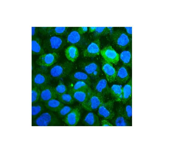
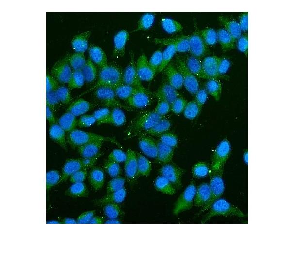
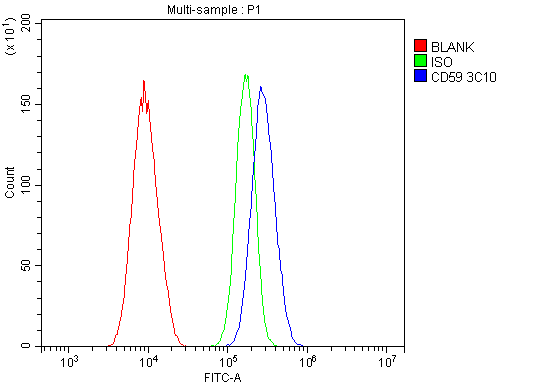
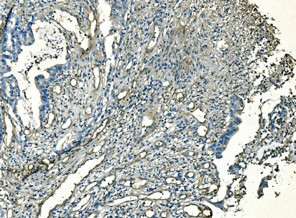
| Application Details | Western blot, 0.1-0.5 µg/ml, Human Immunohistochemistry (Paraffin-embedded Section), 2-5 µg/ml, Human Immunocytochemistry/Immunofluorescence, 2 µg/ml, Human Flow Cytometry, 1-3 µg/1x10^6, Human |
| Protein Name | CD59 molecule, complement regulatory protein |
| Contents | Each vial contains 4mg Trehalose, 0.9mg NaCl, 0.2mg Na2HPO4, 0.05mg NaN3. |
| Clone Names | Clone: 3C10 |
| Immunogen | E.coli-derived human CD59 recombinant protein (Position: L26-N102). Human CD59 shares 47.1% amino acid (aa) sequence identity with rat CD59. |
| Purification | Immunogen affinity purified. |
| Cross Reactivity | No cross-reactivity with other proteins. |
| Storage | Store at -20˚C for one year from date of receipt. After reconstitution, at 4˚C for one month. It can also be aliquotted and stored frozen at -20˚C for six months. Avoid repeated freeze-thaw cycles. |
| Name | CD59 |
|---|---|
| Synonyms | MIC11, MIN1, MIN2, MIN3, MSK21 |
| Function | Potent inhibitor of the complement membrane attack complex (MAC) action. Acts by binding to the C8 and/or C9 complements of the assembling MAC, thereby preventing incorporation of the multiple copies of C9 required for complete formation of the osmolytic pore. This inhibitor appears to be species-specific. Involved in signal transduction for T-cell activation complexed to a protein tyrosine kinase. |
| Cellular Location | Cell membrane; Lipid-anchor, GPI-anchor. Secreted. Note=Soluble form found in a number of tissues |

Thousands of laboratories across the world have published research that depended on the performance of antibodies from Abcepta to advance their research. Check out links to articles that cite our products in major peer-reviewed journals, organized by research category.
info@abcepta.com, and receive a free "I Love Antibodies" mug.

Provided below are standard protocols that you may find useful for product applications.
Background
This gene encodes a cell surface glycoprotein that regulates complement-mediated cell lysis, and it is involved in lymphocyte signal transduction. And this protein is a potent inhibitor of the complement membrane attack complex, whereby it binds complement C8 and/or C9 during the assembly of this complex, thereby inhibiting the incorporation of multiple copies of C9 into the complex, which is necessary for osmolytic pore formation. It also plays a role in signal transduction pathways in the activation of T cells. Mutations in this gene cause CD59 deficiency, a disease resulting in hemolytic anemia and thrombosis, and which causes cerebral infarction. Multiple alternatively spliced transcript variants, which encode the same protein, have been identified for this gene.
If you have used an Abcepta product and would like to share how it has performed, please click on the "Submit Review" button and provide the requested information. Our staff will examine and post your review and contact you if needed.
If you have any additional inquiries please email technical services at tech@abcepta.com.
Ordering Information
Other Products
Shipping Information
