


 Foundational characteristics of cancer include proliferation, angiogenesis, migration, evasion of apoptosis, and cellular immortality. Find key markers for these cellular processes and antibodies to detect them.
 The SUMOplot™ Analysis Program predicts and scores sumoylation sites in your protein. SUMOylation is a post-translational modification involved in various cellular processes, such as nuclear-cytosolic transport, transcriptional regulation, apoptosis, protein stability, response to stress, and progression through the cell cycle.
 The Autophagy Receptor Motif Plotter predicts and scores autophagy receptor binding sites in your protein. Identifying proteins connected to this pathway is critical to understanding the role of autophagy in physiological as well as pathological processes such as development, differentiation, neurodegenerative diseases, stress, infection, and cancer.
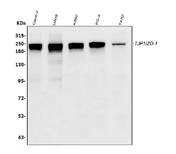
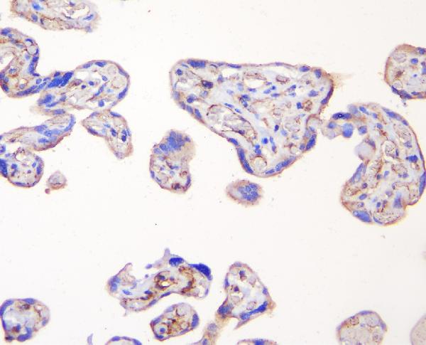
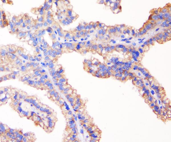
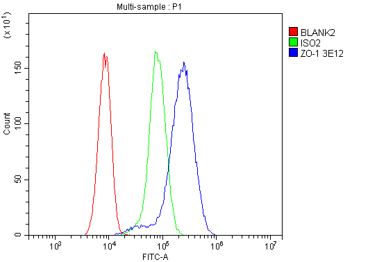
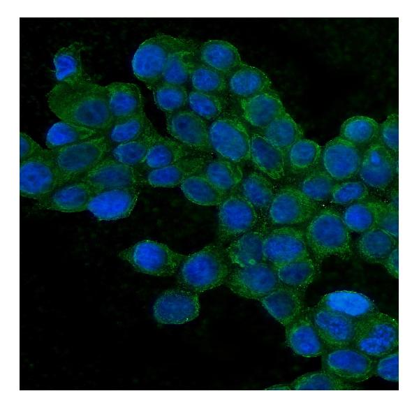
Anti-TJP1 Antibody Picoband™ (monoclonal, 3E12)
- SPECIFICATION
- CITATIONS
- PROTOCOLS
- BACKGROUND

Application
| WB, IHC, IF, ICC, FC |
|---|---|
| Primary Accession | Q07157 |
| Host | Mouse |
| Isotype | Mouse IgG1 |
| Reactivity | Human |
| Clonality | Monoclonal |
| Format | Lyophilized |
| Description | Anti-TJP1 Antibody Picoband™ (monoclonal, 3E12) . Tested in Flow Cytometry, IF, IHC, ICC, WB applications. This antibody reacts with Human. |
| Reconstitution | Add 0.2ml of distilled water will yield a concentration of 500 µg/ml. |
| Gene ID | 7082 |
|---|---|
| Other Names | Tight junction protein ZO-1, Tight junction protein 1, Zona occludens protein 1, Zonula occludens protein 1, TJP1, ZO1 |
| Calculated MW | 220 kDa |
| Application Details | Western blot, 0.1-0.5 µg/ml, Human Immunohistochemistry (Paraffin-embedded Section), 0.5-1 µg/ml, Human, By Heat Immunocytochemistry/Immunofluorescence, 2 µg/ml, Human Flow Cytometry, 1-3 µg/1x10^6 cells, Human |
| Subcellular Localization | Cell membrane; Peripheral membrane protein; Cytoplasmic side; tight junction; Cell junction; gap junction; podosome |
| Tissue Specificity | The alpha-containing isoform is found in most epithelial cell junctions. The short isoform is found both in endothelial cells and the highly specialized epithelial junctions of renal glomeruli and Sertoli cells of the seminiferous tubules. |
| Contents | Each vial contains 4mg Trehalose, 0.9mg NaCl, 0.2mg Na2HPO4, 0.05mg NaN3. |
| Clone Names | Clone: 3E12 |
| Immunogen | E.coli-derived human TJP1 recombinant protein (Position: H1178-F1527). Human TJP1 shares 82% amino acid (aa) sequence identity with mouse TJP1. |
| Cross Reactivity | No cross-reactivity with other proteins. |
| Storage | Store at -20˚C for one year from date of receipt. After reconstitution, at 4˚C for one month. It can also be aliquotted and stored frozen at -20˚C for six months. Avoid repeated freeze-thaw cycles. |
| Name | TJP1 |
|---|---|
| Synonyms | ZO1 |
| Function | TJP1, TJP2, and TJP3 are closely related scaffolding proteins that link tight junction (TJ) transmembrane proteins such as claudins, junctional adhesion molecules, and occludin to the actin cytoskeleton (PubMed:7798316, PubMed:9792688). The tight junction acts to limit movement of substances through the paracellular space and as a boundary between the compositionally distinct apical and basolateral plasma membrane domains of epithelial and endothelial cells. Necessary for lumenogenesis, and particularly efficient epithelial polarization and barrier formation (By similarity). Plays a role in the regulation of cell migration by targeting CDC42BPB to the leading edge of migrating cells (PubMed:21240187). Plays an important role in podosome formation and associated function, thus regulating cell adhesion and matrix remodeling (PubMed:20930113). With TJP2 and TJP3, participates in the junctional retention and stability of the transcription factor DBPA, but is not involved in its shuttling to the nucleus (By similarity). |
| Cellular Location | Cell membrane; Peripheral membrane protein; Cytoplasmic side. Cell junction, tight junction. Cell junction. Cell junction, gap junction. Cell projection, podosome. Note=Moves from the cytoplasm to the cell membrane concurrently with cell-cell contact (PubMed:7798316). At podosomal sites, is predominantly localized in the ring structure surrounding the actin core (PubMed:20930113) Colocalizes with SPEF1 at sites of cell-cell contact in intestinal epithelial cells (PubMed:31473225). |
| Tissue Location | The alpha-containing isoform is found in most epithelial cell junctions. The short isoform is found both in endothelial cells and the highly specialized epithelial junctions of renal glomeruli and Sertoli cells of the seminiferous tubules |
Citations (0)

Thousands of laboratories across the world have published research that depended on the performance of antibodies from Abcepta to advance their research. Check out links to articles that cite our products in major peer-reviewed journals, organized by research category.
Submit your citation using an Abcepta antibody to
info@abcepta.com, and receive a free "I Love Antibodies" mug.
info@abcepta.com, and receive a free "I Love Antibodies" mug.

Application Protocols
Provided below are standard protocols that you may find useful for product applications.
Abcepta welcomes feedback from its customers.
If you have used an Abcepta product and would like to share how it has performed, please click on the "Submit Review" button and provide the requested information. Our staff will examine and post your review and contact you if needed.
If you have any additional inquiries please email technical services at tech@abcepta.com.
$ 370.00
Cat# ABO14891
Ordering Information
United States
AlbaniaAustraliaAustriaBelgiumBosnia & HerzegovinaBrazilBulgariaCanadaCentral AmericaChinaCroatiaCyprusCzech RepublicDenmarkEstoniaFinlandFranceGermanyGreeceHong KongHungaryIcelandIndiaIndonesiaIrelandIsraelItalyJapanLatviaLithuaniaLuxembourgMacedoniaMalaysiaMaltaNetherlandsNew ZealandNorwayPakistanPolandPortugalRomaniaSerbiaSingaporeSlovakiaSloveniaSouth AfricaSouth KoreaSpainSwedenSwitzerlandTaiwanTurkeyUnited KingdomUnited StatesVietnamWorldwideOthers
USA Headquarters
(888) 735-7227 / (858) 622-0099 or (858) 875-1900
Other Products
Shipping Information
Domestic orders (in stock items)
Shipped out the same day. Orders placed after 1 PM (PST) will ship out the next business day.
International orders
Contact your local distributors
