


 Foundational characteristics of cancer include proliferation, angiogenesis, migration, evasion of apoptosis, and cellular immortality. Find key markers for these cellular processes and antibodies to detect them.
 The SUMOplot™ Analysis Program predicts and scores sumoylation sites in your protein. SUMOylation is a post-translational modification involved in various cellular processes, such as nuclear-cytosolic transport, transcriptional regulation, apoptosis, protein stability, response to stress, and progression through the cell cycle.
 The Autophagy Receptor Motif Plotter predicts and scores autophagy receptor binding sites in your protein. Identifying proteins connected to this pathway is critical to understanding the role of autophagy in physiological as well as pathological processes such as development, differentiation, neurodegenerative diseases, stress, infection, and cancer.
Anti-MVP Antibody Picoband™ (monoclonal, 8B12)
- SPECIFICATION
- CITATIONS
- PROTOCOLS
- BACKGROUND

Application
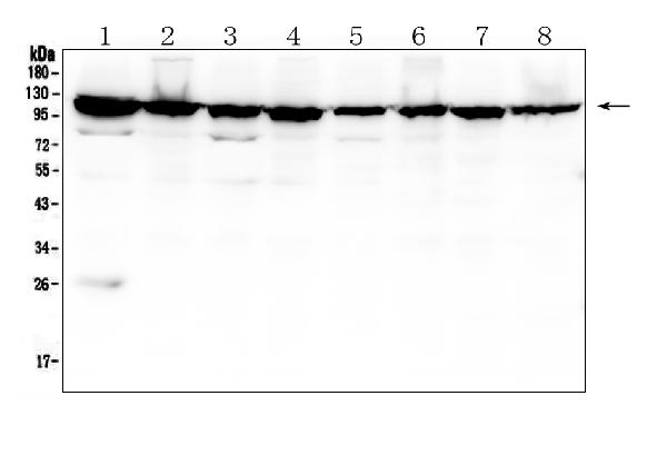
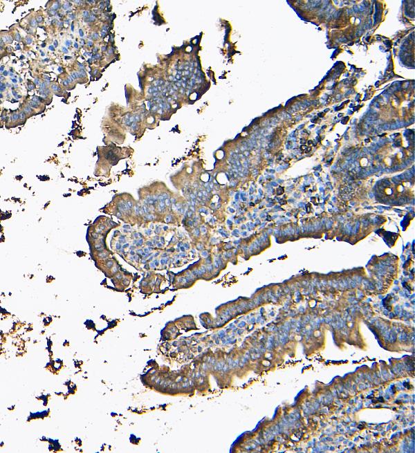
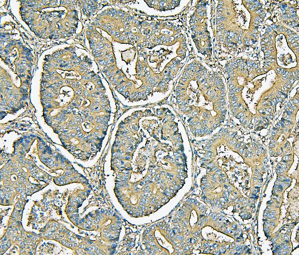
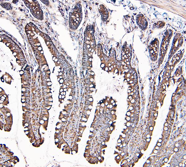
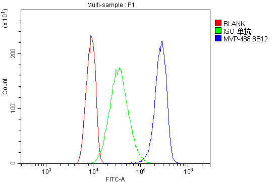
| WB, IHC, IF, ICC, FC |
|---|---|
| Primary Accession | Q14764 |
| Host | Mouse |
| Isotype | Mouse IgG2a |
| Reactivity | Rat, Human, Mouse |
| Clonality | Monoclonal |
| Format | Lyophilized |
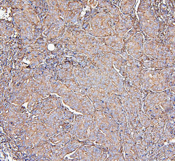
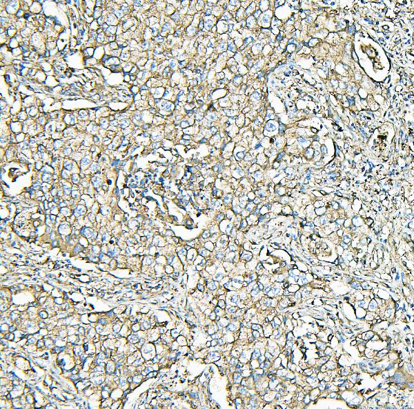
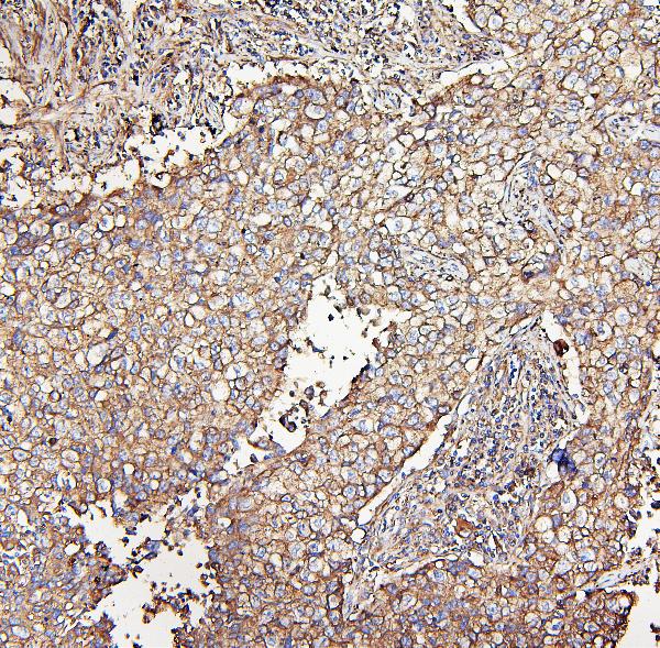
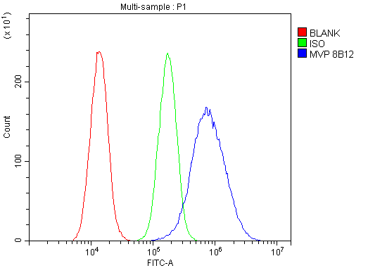
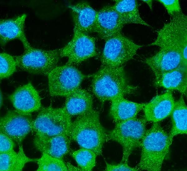
| Description | Anti-MVP Antibody Picoband™ (monoclonal, 8B12) . Tested in Flow Cytometry, IF, IHC, ICC, WB applications. This antibody reacts with Human, Mouse, Rat. |
| Reconstitution | Add 0.2ml of distilled water will yield a concentration of 500 µg/ml. |
| Gene ID | 9961 |
|---|---|
| Other Names | Major vault protein, MVP, Lung resistance-related protein, MVP, LRP |
| Calculated MW | 100-110 kDa |
| Application Details | Western blot, 0.1-0.5 µg/ml, Human, Mouse, Rat Immunohistochemistry (Paraffin-embedded Section), 0.5-1 µg/ml, Human, Mouse, Rat, By Heat Immunocytochemistry/Immunofluorescence, 5 µg/ml, Human Flow Cytometry, 1-3 µg/1x10^6 cells, Human |
| Subcellular Localization | nuclear pore complex; Cytoplasm; perinuclear region |
| Tissue Specificity | Present in most normal tissues. Higher expression observed in epithelial cells with secretory and excretory functions, as well as in cells chronically exposed to xenobiotics, such as bronchial cells and cells lining the intestine. Overexpressed in many multidrug-resistant cancer cells. |
| Contents | Each vial contains 4mg Trehalose, 0.9mg NaCl, 0.2mg Na2HPO4, 0.05mg NaN3. |
| Clone Names | Clone: 8B12 |
| Immunogen | E.coli-derived human MVP recombinant protein (Position: A2-H259). |
| Cross Reactivity | No cross-reactivity with other proteins. |
| Storage | Store at -20˚C for one year from date of receipt. After reconstitution, at 4˚C for one month. It can also be aliquotted and stored frozen at -20˚C for six months. Avoid repeated freeze-thaw cycles. |
| Name | MVP |
|---|---|
| Synonyms | LRP |
| Function | Required for normal vault structure. Vaults are multi-subunit structures that may act as scaffolds for proteins involved in signal transduction. Vaults may also play a role in nucleo-cytoplasmic transport. Down-regulates IFNG-mediated STAT1 signaling and subsequent activation of JAK. Down-regulates SRC activity and signaling through MAP kinases. |
| Cellular Location | Cytoplasm. Nucleus, nuclear pore complex. Cytoplasm, perinuclear region. Note=5% found in the nuclear pore complex (PubMed:15133037). Translocates from the nucleus to the cytoplasm upon EGF treatment (PubMed:16441665) |
| Tissue Location | Present in most normal tissues. Higher expression observed in epithelial cells with secretory and excretory functions, as well as in cells chronically exposed to xenobiotics, such as bronchial cells and cells lining the intestine. Overexpressed in many multidrug- resistant cancer cells |

Thousands of laboratories across the world have published research that depended on the performance of antibodies from Abcepta to advance their research. Check out links to articles that cite our products in major peer-reviewed journals, organized by research category.
info@abcepta.com, and receive a free "I Love Antibodies" mug.

Provided below are standard protocols that you may find useful for product applications.
Background
Major vault protein is a protein that in humans is encoded by the MVP gene. This gene encodes the major component of the vault complex. Vaults are multi-subunit ribonucleoprotein structures that may be involved in nucleo-cytoplasmic transport. The encoded protein may play a role in multiple cellular processes by regulating the MAP kinase, JAK/STAT and phosphoinositide 3-kinase/Akt signaling pathways. The encoded protein also plays a role in multidrug resistance, and expression of this gene may be a prognostic marker for several types of cancer. Alternatively spliced transcript variants have been observed for this gene.
If you have used an Abcepta product and would like to share how it has performed, please click on the "Submit Review" button and provide the requested information. Our staff will examine and post your review and contact you if needed.
If you have any additional inquiries please email technical services at tech@abcepta.com.
Ordering Information
Other Products
Shipping Information
