


 Foundational characteristics of cancer include proliferation, angiogenesis, migration, evasion of apoptosis, and cellular immortality. Find key markers for these cellular processes and antibodies to detect them.
 The SUMOplot™ Analysis Program predicts and scores sumoylation sites in your protein. SUMOylation is a post-translational modification involved in various cellular processes, such as nuclear-cytosolic transport, transcriptional regulation, apoptosis, protein stability, response to stress, and progression through the cell cycle.
 The Autophagy Receptor Motif Plotter predicts and scores autophagy receptor binding sites in your protein. Identifying proteins connected to this pathway is critical to understanding the role of autophagy in physiological as well as pathological processes such as development, differentiation, neurodegenerative diseases, stress, infection, and cancer.
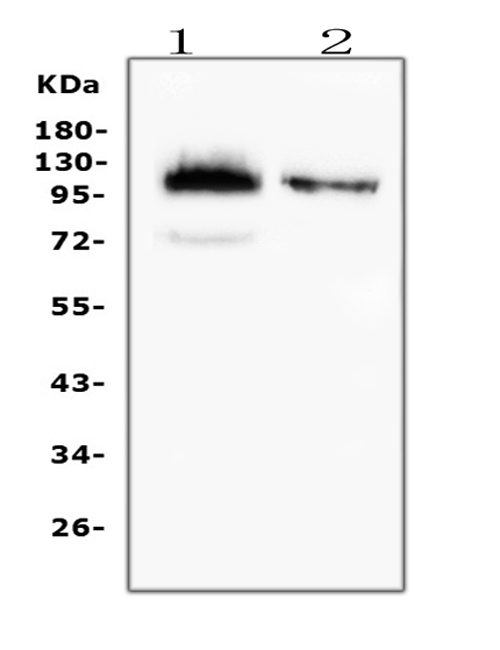
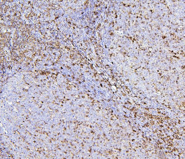
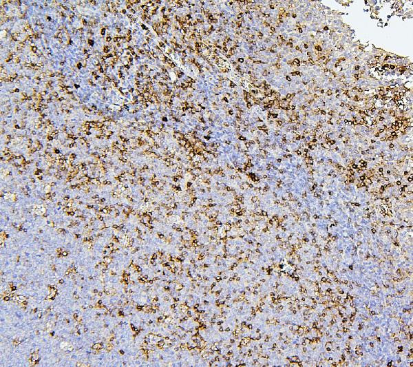
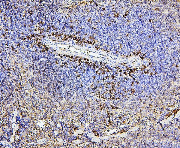
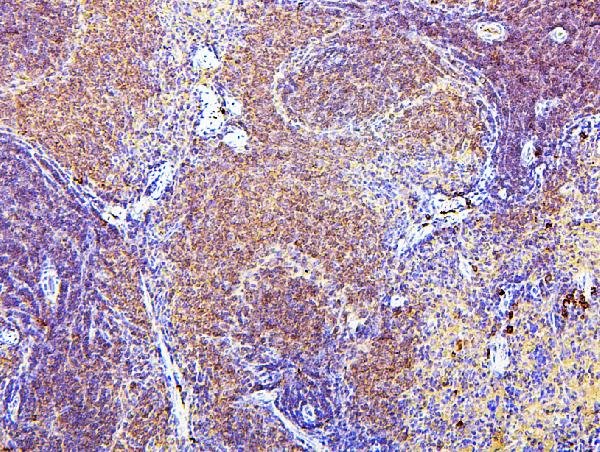
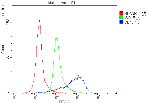
Anti-CD43/SPN Antibody Picoband™ (monoclonal, 4I3)

- SPECIFICATION
- CITATIONS
- PROTOCOLS
- BACKGROUND

Application
| WB, IHC, FC |
|---|---|
| Primary Accession | P16150 |
| Host | Mouse |
| Isotype | Mouse IgG2b |
| Reactivity | Rat, Human, Mouse |
| Clonality | Monoclonal |
| Format | Lyophilized |
| Description | Anti-CD43/SPN Antibody Picoband™ (monoclonal, 4I3) . Tested in Flow Cytometry, IHC, WB applications. This antibody reacts with Human, Mouse, Rat. |
| Reconstitution | Add 0.2ml of distilled water will yield a concentration of 500 µg/ml. |
| Gene ID | 6693 |
|---|---|
| Other Names | Leukosialin, GPL115, Galactoglycoprotein, GALGP, Leukocyte sialoglycoprotein, Sialophorin, CD43, CD43 cytoplasmic tail, CD43-ct, CD43ct, SPN, CD43 |
| Calculated MW | 115 kDa |
| Application Details | Western blot, 0.1-0.5 µg/ml, Human Immunohistochemistry (Paraffin-embedded Section), 0.5-1 µg/ml, Human, Mouse, Rat, By Heat Flow Cytometry, 1-3 µg/1x10^6 cells, Human |
| Subcellular Localization | Membrane. Single-pass type I membrane protein. Microvillus. Uropodium. Nucleus. PML body. |
| Tissue Specificity | Cell surface of thymocytes, T-lymphocytes, neutrophils, plasma cells and myelomas. |
| Contents | Each vial contains 4mg Trehalose, 0.9mg NaCl, 0.2mg Na2HPO4, 0.05mg NaN3. |
| Clone Names | Clone: 4I3 |
| Immunogen | E.coli-derived human CD43 recombinant protein (Position: A272-P400). Human CD43 shares 72% and 73% amino acid (aa) sequence identity with mouse and rat CD43, respectively. |
| Cross Reactivity | No cross-reactivity with other proteins. |
| Storage | Store at -20˚C for one year from date of receipt. After reconstitution, at 4˚C for one month. It can also be aliquotted and stored frozen at -20˚C for six months. Avoid repeated freeze-thaw cycles. |
| Name | SPN |
|---|---|
| Synonyms | CD43 |
| Function | Predominant cell surface sialoprotein of leukocytes which regulates multiple T-cell functions, including T-cell activation, proliferation, differentiation, trafficking and migration. Positively regulates T-cell trafficking to lymph-nodes via its association with ERM proteins (EZR, RDX and MSN) (By similarity). Negatively regulates Th2 cell differentiation and predisposes the differentiation of T-cells towards a Th1 lineage commitment. Promotes the expression of IFN-gamma by T-cells during T-cell receptor (TCR) activation of naive cells and induces the expression of IFN-gamma by CD4(+) T-cells and to a lesser extent by CD8(+) T-cells (PubMed:18036228). Plays a role in preparing T-cells for cytokine sensing and differentiation into effector cells by inducing the expression of cytokine receptors IFNGR and IL4R, promoting IFNGR and IL4R signaling and by mediating the clustering of IFNGR with TCR (PubMed:24328034). Acts as a major E-selectin ligand responsible for Th17 cell rolling on activated vasculature and recruitment during inflammation. Mediates Th17 cells, but not Th1 cells, adhesion to E- selectin. Acts as a T-cell counter-receptor for SIGLEC1 (By similarity). |
| Cellular Location | Membrane; Single-pass type I membrane protein. Cell projection, microvillus {ECO:0000250|UniProtKB:P13838}. Cell projection, uropodium {ECO:0000250|UniProtKB:P15702}. Note=Localizes to the uropodium and microvilli via its interaction with ERM proteins (EZR, RDX and MSN) {ECO:0000250|UniProtKB:P13838, ECO:0000250|UniProtKB:P15702} |
| Tissue Location | Cell surface of thymocytes, T-lymphocytes, neutrophils, plasma cells and myelomas |

Thousands of laboratories across the world have published research that depended on the performance of antibodies from Abcepta to advance their research. Check out links to articles that cite our products in major peer-reviewed journals, organized by research category.
info@abcepta.com, and receive a free "I Love Antibodies" mug.

Provided below are standard protocols that you may find useful for product applications.
Background
CD43, also known as leukosialin or sialophorin, is a transmembrane cell surface protein that in humans is encoded by the SPN gene. It is mapped to 16p11.2. It is a major sialoglycoprotein on the surface of human T lymphocytes, monocytes, granulocytes, and some B lymphocytes, which is important for immune function and may be part of a physiologic ligand-receptor complex involved in T-cell activation. Expression of CD43 is deficient and/or defective in the X-chromosome-linked immunodeficiency disorder Wiscott-Aldrich syndrome, suggesting that CD43 have a role in T-cell activation. T-cell activation requires the removal of CD43 from the immunologic synapse to allow efficient engagement of the TCR with molecules on the APC.
If you have used an Abcepta product and would like to share how it has performed, please click on the "Submit Review" button and provide the requested information. Our staff will examine and post your review and contact you if needed.
If you have any additional inquiries please email technical services at tech@abcepta.com.
Ordering Information
Other Products
Shipping Information
