


 Foundational characteristics of cancer include proliferation, angiogenesis, migration, evasion of apoptosis, and cellular immortality. Find key markers for these cellular processes and antibodies to detect them.
 The SUMOplot™ Analysis Program predicts and scores sumoylation sites in your protein. SUMOylation is a post-translational modification involved in various cellular processes, such as nuclear-cytosolic transport, transcriptional regulation, apoptosis, protein stability, response to stress, and progression through the cell cycle.
 The Autophagy Receptor Motif Plotter predicts and scores autophagy receptor binding sites in your protein. Identifying proteins connected to this pathway is critical to understanding the role of autophagy in physiological as well as pathological processes such as development, differentiation, neurodegenerative diseases, stress, infection, and cancer.
> home > Products > Primary Antibodies > Stem Cells > Anti-Desmin Antibody Picoband™ (monoclonal, 2B5)
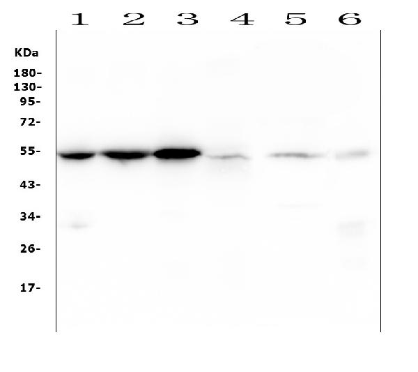
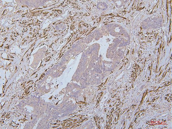
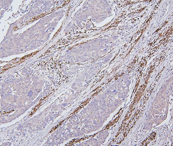
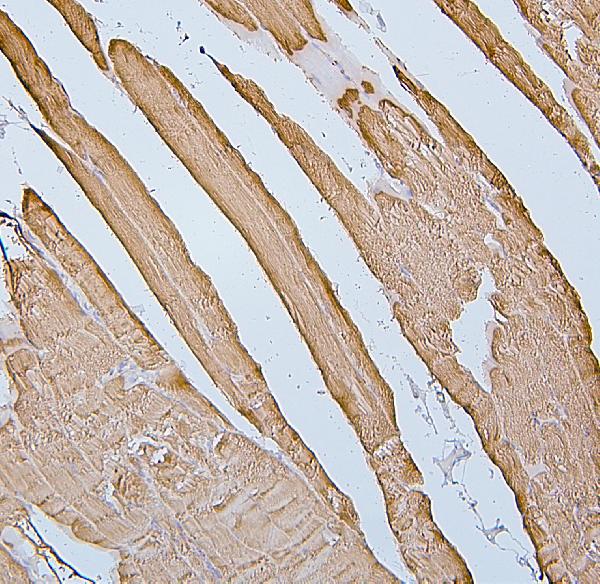
Anti-Desmin Antibody Picoband™ (monoclonal, 2B5)
- SPECIFICATION
- CITATIONS
- PROTOCOLS
- BACKGROUND

Application
| WB, IHC, FC |
|---|---|
| Primary Accession | P17661 |
| Host | Mouse |
| Isotype | Mouse IgG2b |
| Reactivity | Rat, Human, Mouse |
| Clonality | Monoclonal |
| Format | Lyophilized |
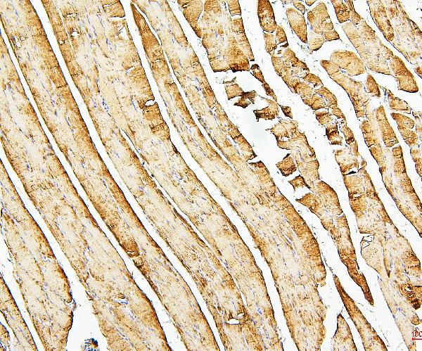
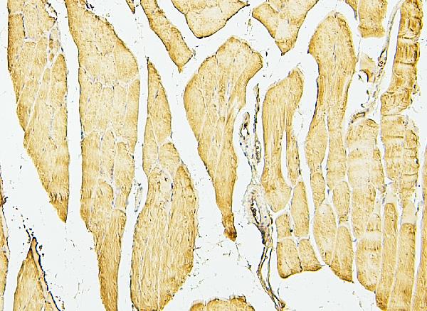
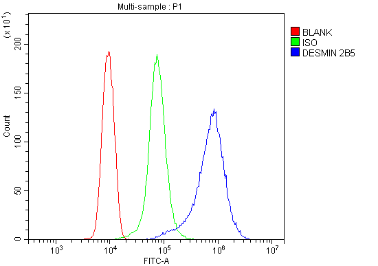
| Description | Anti-Desmin Antibody Picoband™ (monoclonal, 2B5) . Tested in Flow Cytometry, IHC, WB applications. This antibody reacts with Human, Mouse, Rat. |
| Reconstitution | Add 0.2ml of distilled water will yield a concentration of 500 µg/ml. |
| Gene ID | 1674 |
|---|---|
| Other Names | Desmin, DES |
| Calculated MW | 54 kDa |
| Application Details | Western blot, 0.1-0.5 µg/ml Immunohistochemistry (Paraffin-embedded Section), 0.5-1 µg/ml Flow Cytometry, 1-3 µg/1x10^6 cells |
| Subcellular Localization | Nucleus. Sarcolemma. Z line. Cytoplasm. |
| Contents | Each vial contains 4mg Trehalose, 0.9mg NaCl, 0.2mg Na2HPO4, 0.05mg NaN3. |
| Clone Names | Clone: 2B5 |
| Immunogen | E.coli-derived human Desmin recombinant protein (Position: M1-T304). Human Desmin shares 97% amino acid (aa) sequence identity with both mouse and rat Desmin. |
| Cross Reactivity | No cross-reactivity with other proteins. |
| Storage | Store at -20˚C for one year from date of receipt. After reconstitution, at 4˚C for one month. It can also be aliquotted and stored frozen at -20˚C for six months. Avoid repeated freeze-thaw cycles. |
| Name | DES |
|---|---|
| Function | Muscle-specific type III intermediate filament essential for proper muscular structure and function. Plays a crucial role in maintaining the structure of sarcomeres, inter-connecting the Z-disks and forming the myofibrils, linking them not only to the sarcolemmal cytoskeleton, but also to the nucleus and mitochondria, thus providing strength for the muscle fiber during activity (PubMed:25358400). In adult striated muscle they form a fibrous network connecting myofibrils to each other and to the plasma membrane from the periphery of the Z- line structures (PubMed:24200904, PubMed:25394388, PubMed:26724190). May act as a sarcomeric microtubule-anchoring protein: specifically associates with detyrosinated tubulin-alpha chains, leading to buckled microtubules and mechanical resistance to contraction. Required for nuclear membrane integrity, via anchoring at the cell tip and nuclear envelope, resulting in maintenance of microtubule-derived intracellular mechanical forces (By similarity). Contributes to the transcriptional regulation of the NKX2-5 gene in cardiac progenitor cells during a short period of cardiomyogenesis and in cardiac side population stem cells in the adult. Plays a role in maintaining an optimal conformation of nebulette (NEB) on heart muscle sarcomeres to bind and recruit cardiac alpha-actin (By similarity). |
| Cellular Location | Cytoplasm, myofibril, sarcomere, Z line. Cytoplasm Cell membrane, sarcolemma. Nucleus {ECO:0000250|UniProtKB:P31001}. Cell tip {ECO:0000250|UniProtKB:P31001}. Nucleus envelope {ECO:0000250|UniProtKB:P31001}. Note=Localizes in the intercalated disks which occur at the Z line of cardiomyocytes (PubMed:24200904, PubMed:26724190). Localizes in the nucleus exclusively in differentiating cardiac progenitor cells and premature cardiomyocytes (By similarity). PKP2 is required for correct anchoring of DES at the cell tip and nuclear envelope (By similarity) {ECO:0000250|UniProtKB:P31001, ECO:0000269|PubMed:24200904, ECO:0000269|PubMed:26724190} |
Research Areas
Citations (0)

Thousands of laboratories across the world have published research that depended on the performance of antibodies from Abcepta to advance their research. Check out links to articles that cite our products in major peer-reviewed journals, organized by research category.
Submit your citation using an Abcepta antibody to
info@abcepta.com, and receive a free "I Love Antibodies" mug.
info@abcepta.com, and receive a free "I Love Antibodies" mug.

Application Protocols
Provided below are standard protocols that you may find useful for product applications.
Abcepta welcomes feedback from its customers.
If you have used an Abcepta product and would like to share how it has performed, please click on the "Submit Review" button and provide the requested information. Our staff will examine and post your review and contact you if needed.
If you have any additional inquiries please email technical services at tech@abcepta.com.
$ 370.00
Cat# ABO14885
Ordering Information
United States
AlbaniaAustraliaAustriaBelgiumBosnia & HerzegovinaBrazilBulgariaCanadaCentral AmericaChinaCroatiaCyprusCzech RepublicDenmarkEstoniaFinlandFranceGermanyGreeceHong KongHungaryIcelandIndiaIndonesiaIrelandIsraelItalyJapanLatviaLithuaniaLuxembourgMacedoniaMalaysiaMaltaNetherlandsNew ZealandNorwayPakistanPolandPortugalRomaniaSerbiaSingaporeSlovakiaSloveniaSouth AfricaSouth KoreaSpainSwedenSwitzerlandTaiwanTurkeyUnited KingdomUnited StatesVietnamWorldwideOthersMexico
USA Headquarters
(888) 735-7227 / (858) 622-0099 or (858) 875-1900
Other Products
Shipping Information
Domestic orders (in stock items)
Shipped out the same day. Orders placed after 1 PM (PST) will ship out the next business day.
International orders
Contact your local distributors
