


 Foundational characteristics of cancer include proliferation, angiogenesis, migration, evasion of apoptosis, and cellular immortality. Find key markers for these cellular processes and antibodies to detect them.
 The SUMOplot™ Analysis Program predicts and scores sumoylation sites in your protein. SUMOylation is a post-translational modification involved in various cellular processes, such as nuclear-cytosolic transport, transcriptional regulation, apoptosis, protein stability, response to stress, and progression through the cell cycle.
 The Autophagy Receptor Motif Plotter predicts and scores autophagy receptor binding sites in your protein. Identifying proteins connected to this pathway is critical to understanding the role of autophagy in physiological as well as pathological processes such as development, differentiation, neurodegenerative diseases, stress, infection, and cancer.
Anti-COPE Antibody Picoband™ (monoclonal, 9B6)
- SPECIFICATION
- CITATIONS
- PROTOCOLS
- BACKGROUND

Application
| WB, IHC, IF, ICC, FC |
|---|---|
| Primary Accession | O14579 |
| Host | Mouse |
| Isotype | Mouse IgG2a |
| Reactivity | Rat, Human, Mouse |
| Clonality | Monoclonal |
| Format | Lyophilized |
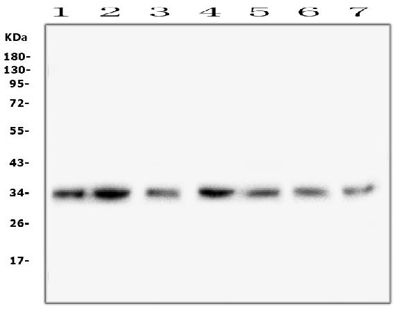
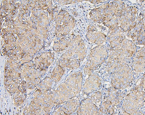
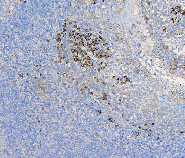
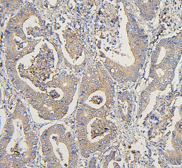
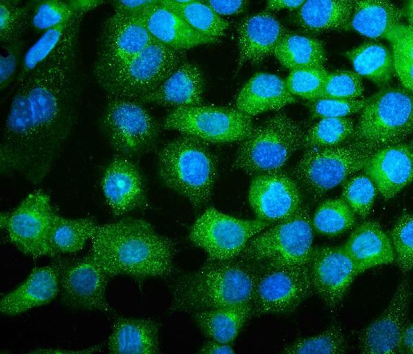
| Description | Anti-COPE Antibody Picoband™ (monoclonal, 9B6) . Tested in Flow Cytometry, IF, IHC, ICC, WB applications. This antibody reacts with Human, Mouse, Rat. |
| Reconstitution | Add 0.2ml of distilled water will yield a concentration of 500 µg/ml. |
| Gene ID | 11316 |
|---|---|
| Other Names | Coatomer subunit epsilon, Epsilon-coat protein, Epsilon-COP, COPE |
| Calculated MW | 34 kDa |
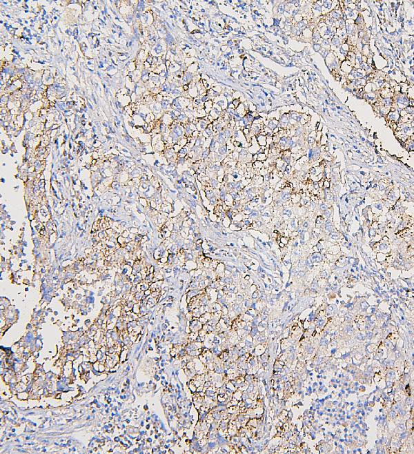
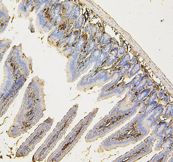
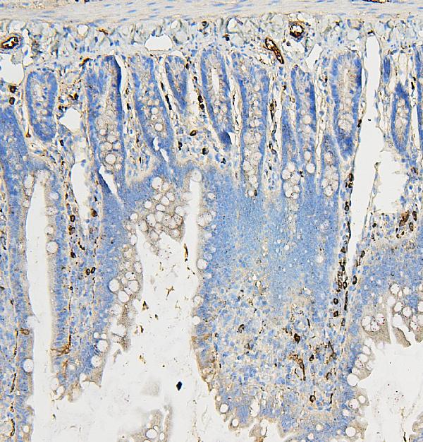
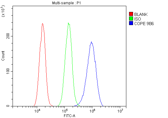
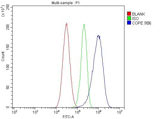
| Application Details | Western blot, 0.1-0.5 µg/ml Immunohistochemistry (Paraffin-embedded Section), 0.5-1 µg/ml Immunocytochemistry/Immunofluorescence, 5 µg/ml Flow Cytometry, 1-3 µg/1x10^6 cells |
| Subcellular Localization | Golgi apparatus membrane. Peripheral membrane protein. Cytoplasmic side. Cytoplasm. COPI-coated vesicle membrane. |
| Contents | Each vial contains 4mg Trehalose, 0.9mg NaCl, 0.2mg Na2HPO4, 0.05mg NaN3. |
| Clone Names | Clone: 9B6 |
| Immunogen | E. coli-derived human COPE recombinant protein (Position: E80-A308). Human COPE shares 89.5% amino acid (aa) sequence identity with mouse COPE. |
| Cross Reactivity | No cross-reactivity with other proteins. |
| Storage | Store at -20˚C for one year from date of receipt. After reconstitution, at 4˚C for one month. It can also be aliquotted and stored frozen at -20˚C for six months. Avoid repeated freeze-thaw cycles. |
| Name | COPE |
|---|---|
| Function | The coatomer is a cytosolic protein complex that binds to dilysine motifs and reversibly associates with Golgi non-clathrin- coated vesicles, which further mediate biosynthetic protein transport from the ER, via the Golgi up to the trans Golgi network. The coatomer complex is required for budding from Golgi membranes, and is essential for the retrograde Golgi-to-ER transport of dilysine-tagged proteins. In mammals, the coatomer can only be recruited by membranes associated with ADP-ribosylation factors (ARFs), which are small GTP-binding proteins; the complex also influences the Golgi structural integrity, as well as the processing, activity, and endocytic recycling of LDL receptors (By similarity). |
| Cellular Location | Cytoplasm. Golgi apparatus membrane; Peripheral membrane protein; Cytoplasmic side. Cytoplasmic vesicle, COPI-coated vesicle membrane; Peripheral membrane protein; Cytoplasmic side. Note=The coatomer is cytoplasmic or polymerized on the cytoplasmic side of the Golgi, as well as on the vesicles/buds originating from it. |

Thousands of laboratories across the world have published research that depended on the performance of antibodies from Abcepta to advance their research. Check out links to articles that cite our products in major peer-reviewed journals, organized by research category.
info@abcepta.com, and receive a free "I Love Antibodies" mug.

Provided below are standard protocols that you may find useful for product applications.
Background
Coatomer subunit epsilon is a protein that in humans is encoded by the COPE gene. The product of this gene is an epsilon subunit of coatomer protein complex. Coatomer is a cytosolic protein complex that binds to dilysine motifs and reversibly associates with Golgi non-clathrin-coated vesicles. It is required for budding from Golgi membranes, and is essential for the retrograde Golgi-to-ER transport of dilysine-tagged proteins. Coatomer complex consists of at least the alpha, beta, beta', gamma, delta, epsilon and zeta subunits. Alternatively spliced transcript variants encoding different isoforms have been identified.
If you have used an Abcepta product and would like to share how it has performed, please click on the "Submit Review" button and provide the requested information. Our staff will examine and post your review and contact you if needed.
If you have any additional inquiries please email technical services at tech@abcepta.com.
Ordering Information
Other Products
Shipping Information
