


 Foundational characteristics of cancer include proliferation, angiogenesis, migration, evasion of apoptosis, and cellular immortality. Find key markers for these cellular processes and antibodies to detect them.
 The SUMOplot™ Analysis Program predicts and scores sumoylation sites in your protein. SUMOylation is a post-translational modification involved in various cellular processes, such as nuclear-cytosolic transport, transcriptional regulation, apoptosis, protein stability, response to stress, and progression through the cell cycle.
 The Autophagy Receptor Motif Plotter predicts and scores autophagy receptor binding sites in your protein. Identifying proteins connected to this pathway is critical to understanding the role of autophagy in physiological as well as pathological processes such as development, differentiation, neurodegenerative diseases, stress, infection, and cancer.
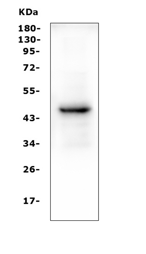
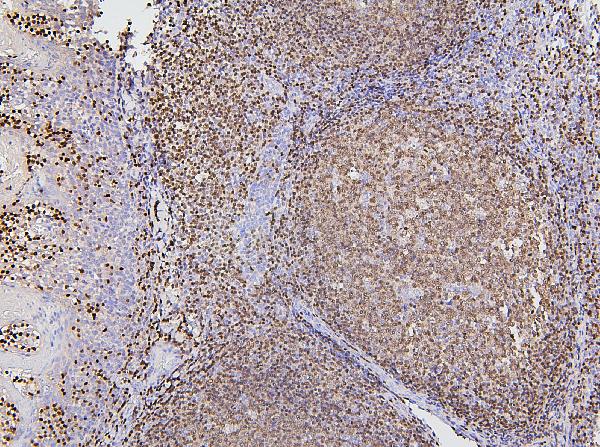
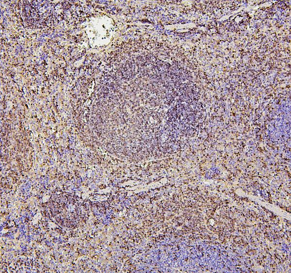
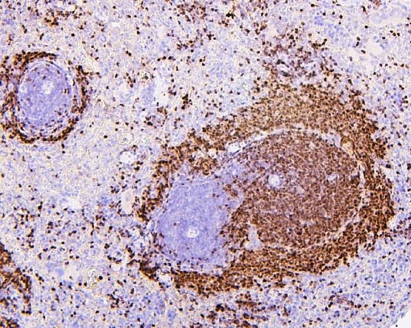
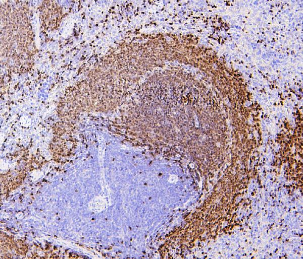
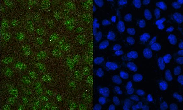
Anti-PAX5 Antibody Picoband™ (monoclonal, 6I4)
- SPECIFICATION
- CITATIONS
- PROTOCOLS
- BACKGROUND

Application
| WB, IHC, IF, ICC |
|---|---|
| Primary Accession | Q02548 |
| Host | Mouse |
| Isotype | Mouse IgG2b |
| Reactivity | Rat, Human, Mouse |
| Clonality | Monoclonal |
| Format | Lyophilized |
| Description | Anti-PAX5 Antibody Picoband™ (monoclonal, 6I4) . Tested in IF, IHC, ICC, WB applications. This antibody reacts with Human, Mouse, Rat. |
| Reconstitution | Add 0.2ml of distilled water will yield a concentration of 500 µg/ml. |
| Gene ID | 5079 |
|---|---|
| Other Names | Paired box protein Pax-5, B-cell-specific transcription factor, BSAP, PAX5 |
| Calculated MW | 45 kDa |
| Application Details | Western blot, 0.1-0.5 µg/ml, Human Immunohistochemistry (Paraffin-embedded Section), 0.5-1 µg/ml, Human, Mouse, Rat, By Heat Immunocytochemistry/Immunofluorescence, 2 µg/ml, Human |
| Subcellular Localization | Nucleus. |
| Contents | Each vial contains 4mg Trehalose, 0.9mg NaCl, 0.2mg Na2HPO4, 0.05mg NaN3. |
| Clone Names | Clone: 6I4 |
| Immunogen | E. coli-derived human PAX5 recombinant protein (Position: R217-A282). |
| Cross Reactivity | No cross-reactivity with other proteins. |
| Storage | Store at -20˚C for one year from date of receipt. After reconstitution, at 4˚C for one month. It can also be aliquotted and stored frozen at -20˚C for six months. Avoid repeated freeze-thaw cycles. |
| Name | PAX5 |
|---|---|
| Function | Transcription factor that plays an essential role in commitment of lymphoid progenitors to the B-lymphocyte lineage (PubMed:10811620, PubMed:27181361). Fulfills a dual role by repressing B-lineage inappropriate genes and simultaneously activating B-lineage- specific genes (PubMed:10811620, PubMed:27181361). In turn, regulates cell adhesion and migration, induces V(H)-to-D(H)J(H) recombination, facilitates pre-B-cell receptor signaling and promotes development to the mature B-cell stage (PubMed:32612238). Repression of the cohesin- release factor WAPL causes global changes of the chromosomal architecture in pro-B cells to facilitate the generation of a diverse antibody repertoire (PubMed:32612238). |
| Cellular Location | Nucleus. |

Thousands of laboratories across the world have published research that depended on the performance of antibodies from Abcepta to advance their research. Check out links to articles that cite our products in major peer-reviewed journals, organized by research category.
info@abcepta.com, and receive a free "I Love Antibodies" mug.

Provided below are standard protocols that you may find useful for product applications.
Background
Paired box protein Pax-5 is a protein that in humans is encoded by the PAX5 gene. The PAX5 gene is a member of the paired box (PAX) family of transcription factors. The central feature of this gene family is a novel, highly conserved DNA-binding domain, known as the paired box. The PAX proteins are important regulators in early development, and alterations in the expression of their genes are thought to contribute to neoplastic transformation. The PAX5 gene encodes the B-cell lineage specific activator protein (BSAP) that is expressed at early, but not late stages of B-cell differentiation. Its expression has also been detected in developing CNS and testis, therefore, PAX5 gene product may not only play an important role in B-cell differentiation, but also in neural development and spermatogenesis.
If you have used an Abcepta product and would like to share how it has performed, please click on the "Submit Review" button and provide the requested information. Our staff will examine and post your review and contact you if needed.
If you have any additional inquiries please email technical services at tech@abcepta.com.
Ordering Information
Other Products
Shipping Information
