


 Foundational characteristics of cancer include proliferation, angiogenesis, migration, evasion of apoptosis, and cellular immortality. Find key markers for these cellular processes and antibodies to detect them.
 The SUMOplot™ Analysis Program predicts and scores sumoylation sites in your protein. SUMOylation is a post-translational modification involved in various cellular processes, such as nuclear-cytosolic transport, transcriptional regulation, apoptosis, protein stability, response to stress, and progression through the cell cycle.
 The Autophagy Receptor Motif Plotter predicts and scores autophagy receptor binding sites in your protein. Identifying proteins connected to this pathway is critical to understanding the role of autophagy in physiological as well as pathological processes such as development, differentiation, neurodegenerative diseases, stress, infection, and cancer.
Anti-Ran Antibody Picoband™ (monoclonal, 5D5)
- SPECIFICATION
- CITATIONS
- PROTOCOLS
- BACKGROUND

Application
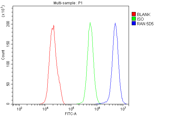
| WB, IF, ICC, FC |
|---|---|
| Primary Accession | P62826 |
| Host | Mouse |
| Isotype | Mouse IgG2b |
| Reactivity | Rat, Human, Mouse |
| Clonality | Monoclonal |
| Format | Lyophilized |
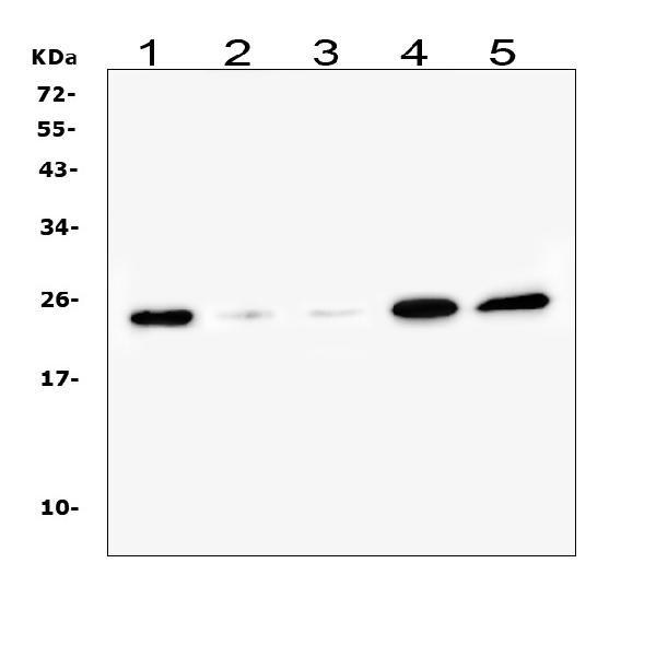
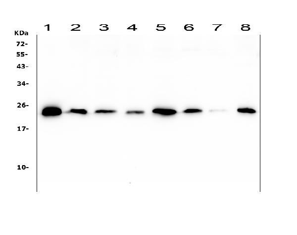
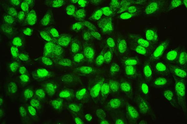
| Description | Anti-Ran Antibody Picoband™ (monoclonal, 5D5) . Tested in Flow Cytometry, IF, ICC, WB applications. This antibody reacts with Human, Mouse, Rat. |
| Reconstitution | Add 0.2ml of distilled water will yield a concentration of 500 µg/ml. |
| Gene ID | 5901 |
|---|---|
| Other Names | GTP-binding nuclear protein Ran, 3.6.5.-, Androgen receptor-associated protein 24, GTPase Ran, Ras-like protein TC4, Ras-related nuclear protein, RAN, ARA24 {ECO:0000303|PubMed:10400640} |
| Calculated MW | 24 kDa |
| Application Details | Western blot, 0.1-0.5 µg/ml Immunocytochemistry/Immunofluorescence, 2 µg/ml Flow Cytometry, 1-3 µg/1x10^6 cells |
| Subcellular Localization | Cytosol. Nucleus. Nucleus envelope. Cytoplasm. Melanosome. |
| Tissue Specificity | Expressed in a variety of tissues. |
| Contents | Each vial contains 4mg Trehalose, 0.9mg NaCl, 0.2mg Na2HPO4, 0.05mg NaN3. |
| Clone Names | Clone: 5D5 |
| Immunogen | E. coli-derived human Ran recombinant protein (Position: A2-L216). Human Ran shares 100% amino acid (aa) sequence identity with both mouse and rat Ran. |
| Cross Reactivity | No cross-reactivity with other proteins. |
| Storage | Store at -20˚C for one year from date of receipt. After reconstitution, at 4˚C for one month. It can also be aliquotted and stored frozen at -20˚C for six months. Avoid repeated freeze-thaw cycles. |
| Name | RAN |
|---|---|
| Synonyms | ARA24 {ECO:0000303|PubMed:10400640} |
| Function | GTPase involved in nucleocytoplasmic transport, participating both to the import and the export from the nucleus of proteins and RNAs (PubMed:10400640, PubMed:17209048, PubMed:26272610, PubMed:27306458, PubMed:8276887, PubMed:8636225, PubMed:8692944, PubMed:8896452, PubMed:9351834, PubMed:9428644, PubMed:9822603). Switches between a cytoplasmic GDP- and a nuclear GTP-bound state by nucleotide exchange and GTP hydrolysis (PubMed:11336674, PubMed:26272610, PubMed:29040603, PubMed:7819259, PubMed:8636225, PubMed:8692944, PubMed:8896452, PubMed:9351834, PubMed:9428644, PubMed:9822603). Nuclear import receptors such as importin beta bind their substrates only in the absence of GTP-bound RAN and release them upon direct interaction with GTP-bound RAN, while export receptors behave in the opposite way. Thereby, RAN controls cargo loading and release by transport receptors in the proper compartment and ensures the directionality of the transport (PubMed:8896452, PubMed:9351834, PubMed:9428644). Interaction with RANBP1 induces a conformation change in the complex formed by XPO1 and RAN that triggers the release of the nuclear export signal of cargo proteins (PubMed:20485264). RAN (GTP-bound form) triggers microtubule assembly at mitotic chromosomes and is required for normal mitotic spindle assembly and chromosome segregation (PubMed:10408446, PubMed:29040603). Required for normal progress through mitosis (PubMed:12194828, PubMed:29040603, PubMed:8421051). The complex with BIRC5/survivin plays a role in mitotic spindle formation by serving as a physical scaffold to help deliver the RAN effector molecule TPX2 to microtubules (PubMed:18591255). Acts as a negative regulator of the kinase activity of VRK1 and VRK2 (PubMed:18617507). Enhances AR- mediated transactivation. Transactivation decreases as the poly-Gln length within AR increases (PubMed:10400640). |
| Cellular Location | Nucleus. Nucleus envelope. Cytoplasm, cytosol Cytoplasm. Melanosome Note=Predominantly nuclear during interphase (PubMed:10679025, PubMed:12194828, PubMed:8421051). Becomes dispersed throughout the cytoplasm during mitosis (PubMed:12194828, PubMed:8421051). Identified by mass spectrometry in melanosome fractions from stage I to stage IV (PubMed:17081065). |
| Tissue Location | Expressed in a variety of tissues. |

Thousands of laboratories across the world have published research that depended on the performance of antibodies from Abcepta to advance their research. Check out links to articles that cite our products in major peer-reviewed journals, organized by research category.
info@abcepta.com, and receive a free "I Love Antibodies" mug.

Provided below are standard protocols that you may find useful for product applications.
Background
RAN (ras-related nuclear protein) is a small GTP binding protein belonging to the RAS superfamily that is essential for the translocation of RNA and proteins through the nuclear pore complex. The RAN protein is also involved in control of DNA synthesis and cell cycle progression. Nuclear localization of RAN requires the presence of regulator of chromosome condensation 1 (RCC1). Mutations in RAN disrupt DNA synthesis. Because of its many functions, it is likely that RAN interacts with several other proteins. RAN regulates formation and organization of the microtubule network independently of its role in the nucleus-cytosol exchange of macromolecules. RAN could be a key signaling molecule regulating microtubule polymerization during mitosis. RCC1 generates a high local concentration of RAN-GTP around chromatin which, in turn, induces the local nucleation of microtubules. RAN is an androgen receptor (AR) coactivator that binds differentially with different lengths of polyglutamine within the androgen receptor. Polyglutamine repeat expansion in the AR is linked to Kennedy's disease (X-linked spinal and bulbar muscular atrophy). RAN coactivation of the AR diminishes with polyglutamine expansion within the AR, and this weak coactivation may lead to partial androgen insensitivity during the development of Kennedy's disease.
If you have used an Abcepta product and would like to share how it has performed, please click on the "Submit Review" button and provide the requested information. Our staff will examine and post your review and contact you if needed.
If you have any additional inquiries please email technical services at tech@abcepta.com.
Ordering Information
Other Products
Shipping Information
