


 Foundational characteristics of cancer include proliferation, angiogenesis, migration, evasion of apoptosis, and cellular immortality. Find key markers for these cellular processes and antibodies to detect them.
 The SUMOplot™ Analysis Program predicts and scores sumoylation sites in your protein. SUMOylation is a post-translational modification involved in various cellular processes, such as nuclear-cytosolic transport, transcriptional regulation, apoptosis, protein stability, response to stress, and progression through the cell cycle.
 The Autophagy Receptor Motif Plotter predicts and scores autophagy receptor binding sites in your protein. Identifying proteins connected to this pathway is critical to understanding the role of autophagy in physiological as well as pathological processes such as development, differentiation, neurodegenerative diseases, stress, infection, and cancer.
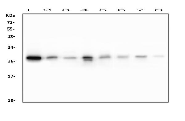
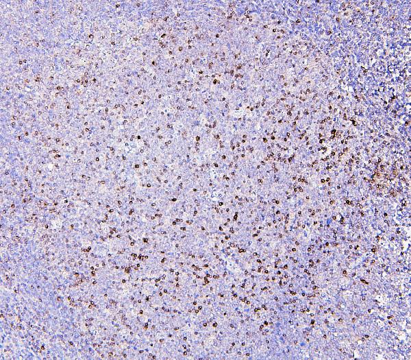
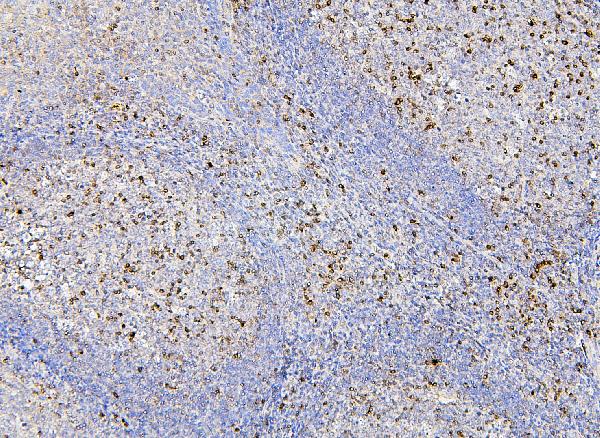
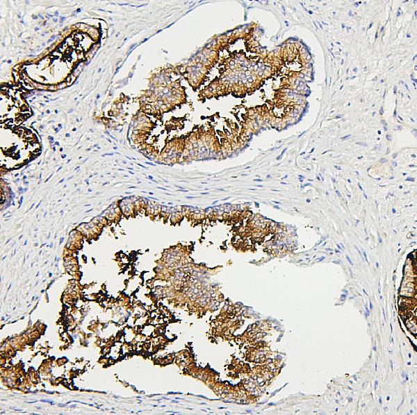
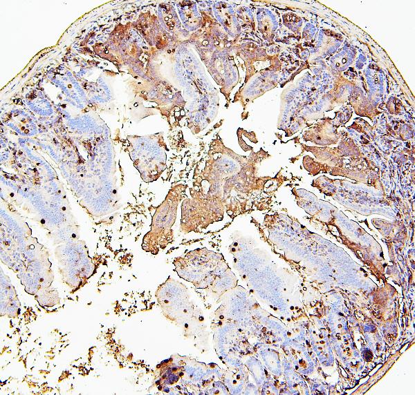
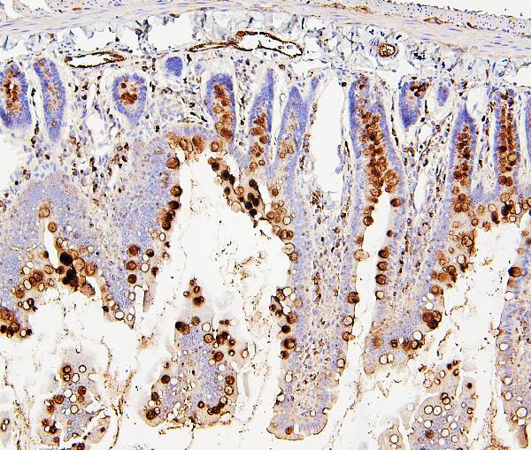
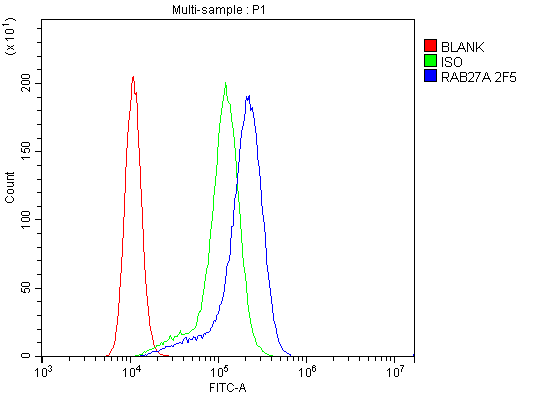
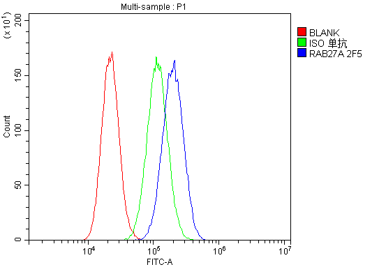
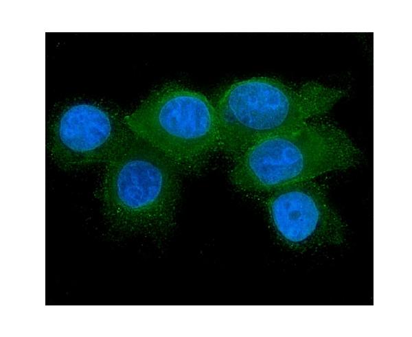
Anti-RAB27A Antibody Picoband™ (monoclonal, 2F5)
- SPECIFICATION
- CITATIONS
- PROTOCOLS
- BACKGROUND

Application
| WB, IHC, IF, ICC, FC |
|---|---|
| Primary Accession | P51159 |
| Host | Mouse |
| Isotype | Mouse IgG2b |
| Reactivity | Rat, Human, Mouse |
| Clonality | Monoclonal |
| Format | Lyophilized |
| Description | Anti-RAB27A Antibody Picoband™ (monoclonal, 2F5) . Tested in Flow Cytometry, IF, IHC, ICC, WB applications. This antibody reacts with Human, Mouse, Rat. |
| Reconstitution | Add 0.2ml of distilled water will yield a concentration of 500 µg/ml. |
| Gene ID | 5873 |
|---|---|
| Other Names | Ras-related protein Rab-27A, Rab-27, 3.6.5.2, GTP-binding protein Ram, RAB27A, RAB27 |
| Calculated MW | 27 kDa |
| Application Details | Western blot, 0.1-0.5 µg/ml Immunohistochemistry (Paraffin-embedded Section), 0.5-1 µg/ml Immunocytochemistry/Immunofluorescence, 2 µg/ml Flow Cytometry, 1-3 µg/1x10^6 cells |
| Subcellular Localization | Lysosome. Late endosome. Membrane. Lipid-anchor. Melanosome. |
| Tissue Specificity | Found in all the examined tissues except in brain. Low expression was found in thymus, kidney, muscle and placenta. Detected in melanocytes, and in most tumor cell lines examined. Expressed in cytotoxic T-lymphocytes (CTL) and mast cells. |
| Contents | Each vial contains 4mg Trehalose, 0.9mg NaCl, 0.2mg Na2HPO4, 0.05mg NaN3. |
| Clone Names | Clone: 2F5 |
| Immunogen | E. coli-derived human RAB27A recombinant protein (Position: L98-K216). |
| Cross Reactivity | No cross-reactivity with other proteins. |
| Storage | Store at -20˚C for one year from date of receipt. After reconstitution, at 4˚C for one month. It can also be aliquotted and stored frozen at -20˚C for six months. Avoid repeated freeze-thaw cycles. |
| Name | RAB27A |
|---|---|
| Synonyms | RAB27 |
| Function | Small GTPase which cycles between active GTP-bound and inactive GDP-bound states. In its active state, binds to a variety of effector proteins to regulate homeostasis of late endocytic pathway, including endosomal positioning, maturation and secretion (PubMed:30771381). Plays a role in cytotoxic granule exocytosis in lymphocytes. Required for both granule maturation and granule docking and priming at the immunologic synapse. |
| Cellular Location | Membrane; Lipid-anchor. Melanosome. Late endosome. Lysosome. Note=Identified by mass spectrometry in melanosome fractions from stage I to stage IV (PubMed:12643545, PubMed:17081065). Localizes to endosomal exocytic vesicles (PubMed:17237785). |
| Tissue Location | Found in all the examined tissues except in brain. Low expression was found in thymus, kidney, muscle and placenta Detected in melanocytes, and in most tumor cell lines examined Expressed in cytotoxic T-lymphocytes (CTL) and mast cells |

Thousands of laboratories across the world have published research that depended on the performance of antibodies from Abcepta to advance their research. Check out links to articles that cite our products in major peer-reviewed journals, organized by research category.
info@abcepta.com, and receive a free "I Love Antibodies" mug.

Provided below are standard protocols that you may find useful for product applications.
Background
Ras-related protein Rab-27A is a protein that in humans is encoded by the RAB27A gene. The protein encoded by this gene belongs to the small GTPase superfamily, Rab family. The protein is membrane-bound and may be involved in protein transport and small GTPase mediated signal transduction. Mutations in this gene are associated with Griscelli syndrome type 2. Alternative splicing occurs at this locus and four transcript variants encoding the same protein have been identified.
If you have used an Abcepta product and would like to share how it has performed, please click on the "Submit Review" button and provide the requested information. Our staff will examine and post your review and contact you if needed.
If you have any additional inquiries please email technical services at tech@abcepta.com.
Ordering Information
Other Products
Shipping Information
