


 Foundational characteristics of cancer include proliferation, angiogenesis, migration, evasion of apoptosis, and cellular immortality. Find key markers for these cellular processes and antibodies to detect them.
 The SUMOplot™ Analysis Program predicts and scores sumoylation sites in your protein. SUMOylation is a post-translational modification involved in various cellular processes, such as nuclear-cytosolic transport, transcriptional regulation, apoptosis, protein stability, response to stress, and progression through the cell cycle.
 The Autophagy Receptor Motif Plotter predicts and scores autophagy receptor binding sites in your protein. Identifying proteins connected to this pathway is critical to understanding the role of autophagy in physiological as well as pathological processes such as development, differentiation, neurodegenerative diseases, stress, infection, and cancer.
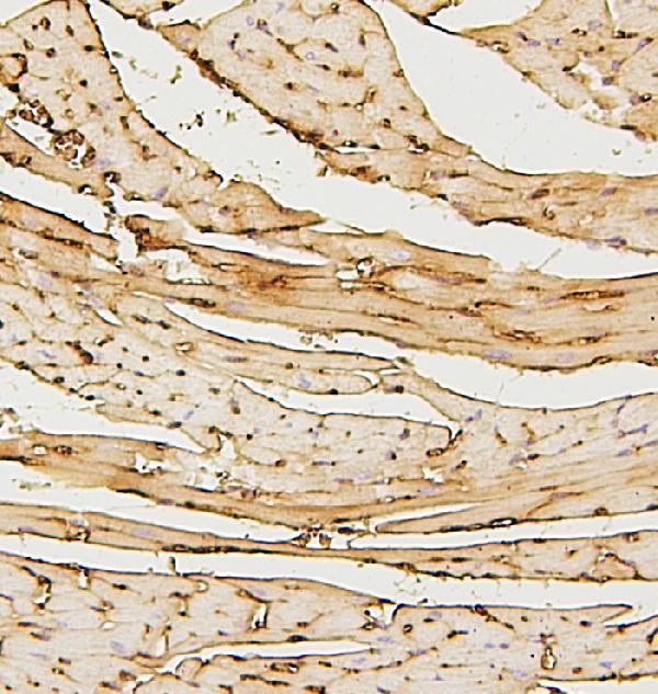
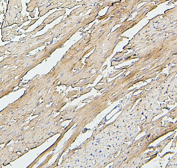
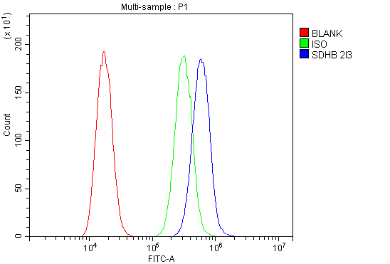
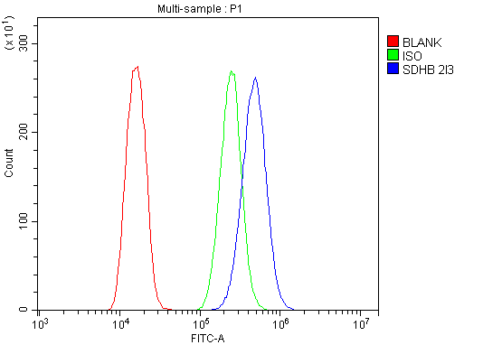
Anti-SDHB Antibody Picoband™ (monoclonal, 2I3)
- SPECIFICATION
- CITATIONS
- PROTOCOLS
- BACKGROUND

Application
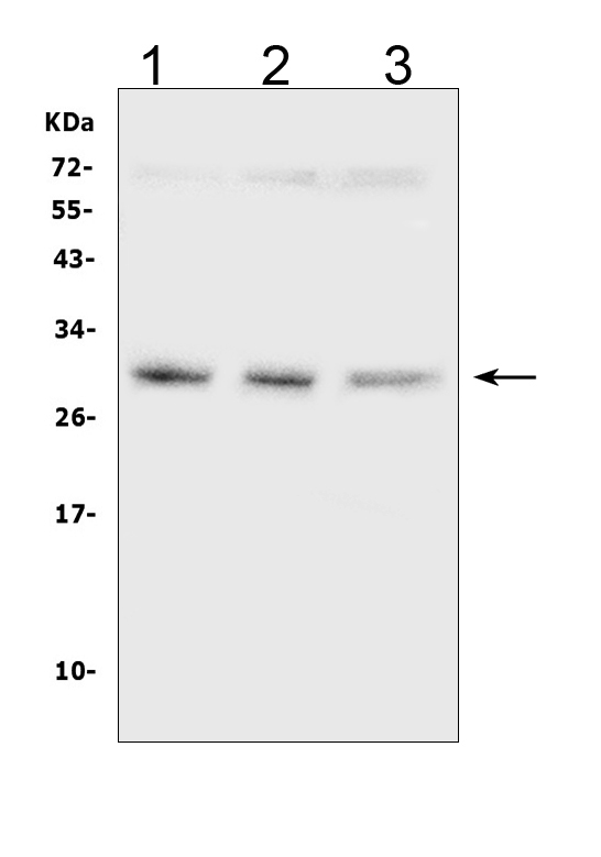
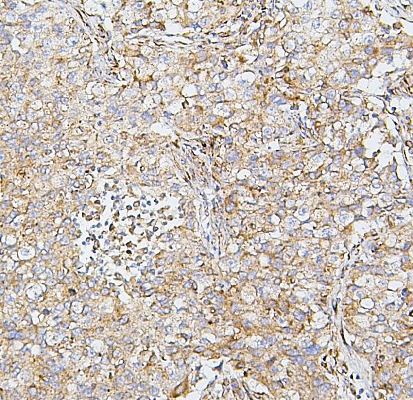
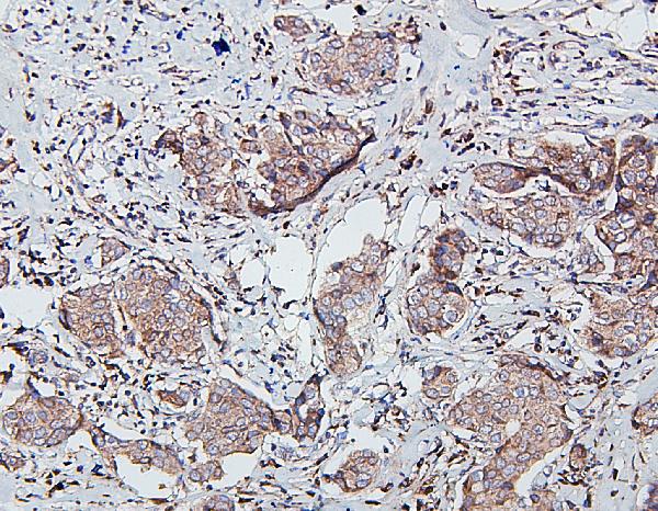
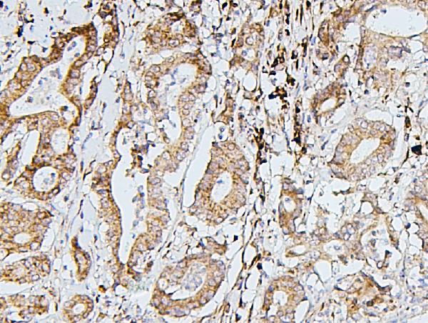
| WB, IHC, FC |
|---|---|
| Primary Accession | P21912 |
| Host | Mouse |
| Isotype | Mouse IgG1 |
| Reactivity | Rat, Human, Mouse |
| Clonality | Monoclonal |
| Format | Lyophilized |
| Description | Anti-SDHB Antibody Picoband™ (monoclonal, 2I3) . Tested in Flow Cytometry, IHC, WB applications. This antibody reacts with Human, Mouse, Rat. |
| Reconstitution | Add 0.2ml of distilled water will yield a concentration of 500 µg/ml. |
| Gene ID | 6390 |
|---|---|
| Other Names | Succinate dehydrogenase [ubiquinone] iron-sulfur subunit, mitochondrial, 1.3.5.1, Iron-sulfur subunit of complex II, Ip, SDHB, SDH, SDH1 |
| Calculated MW | 29 kDa |
| Application Details | Western blot, 0.1-0.5 µg/ml, Human Immunohistochemistry (Paraffin-embedded Section), 0.5-1 µg/ml, Human, Mouse, Rat Flow Cytometry, 1-3 µg/1x10^6 cells, Human |
| Subcellular Localization | Mitochondrion inner membrane. Peripheral membrane protein. Matrix side. |
| Contents | Each vial contains 4mg Trehalose, 0.9mg NaCl, 0.2mg Na2HPO4, 0.05mg NaN3. |
| Clone Names | Clone: 2I3 |
| Immunogen | E. coli-derived human SDHB recombinant protein (Position: A29-V280). |
| Cross Reactivity | No cross-reactivity with other proteins. |
| Storage | Store at -20˚C for one year from date of receipt. After reconstitution, at 4˚C for one month. It can also be aliquotted and stored frozen at -20˚C for six months. Avoid repeated freeze-thaw cycles. |
| Name | SDHB |
|---|---|
| Synonyms | SDH, SDH1 |
| Function | Iron-sulfur protein (IP) subunit of the succinate dehydrogenase complex (mitochondrial respiratory chain complex II), responsible for transferring electrons from succinate to ubiquinone (coenzyme Q) (PubMed:26925370, PubMed:27604842). SDH also oxidizes malate to the non-canonical enol form of oxaloacetate, enol- oxaloacetate (By similarity). Enol-oxaloacetate, which is a potent inhibitor of the succinate dehydrogenase activity, is further isomerized into keto-oxaloacetate (By similarity). |
| Cellular Location | Mitochondrion inner membrane; Peripheral membrane protein; Matrix side |

Thousands of laboratories across the world have published research that depended on the performance of antibodies from Abcepta to advance their research. Check out links to articles that cite our products in major peer-reviewed journals, organized by research category.
info@abcepta.com, and receive a free "I Love Antibodies" mug.

Provided below are standard protocols that you may find useful for product applications.
Background
SDHB (Succinate Dehydrogenase Complex, Subunit B, iron sulfur protein), also known as iron-sulfur subunit of complex II (Ip) or SDH2, HOMOLOG OF, is a protein that in humans is encoded by the SDHB gene. SDHB is one of four protein subunits forming succinate dehydrogenase, the other three being SDHA, SDHC and SDHD. The SDHB subunit is connected to the SDHA subunit on the hydrophilic, catalytic end of the SDH complex. The SDHB gene is mapped on 1p36.13. It is stated that the nuclear-encoded Krebs cycle enzymes fumarate hydratase and succinate dehydrogenases like SDHB act as tumor suppressors, and germline mutations in these genes predispose individuals to leiomyomas and renal cancer and to paragangliomas, respectively. In affected members of families with paragangliomas-4, mutations were identified in the SDHB gene.
If you have used an Abcepta product and would like to share how it has performed, please click on the "Submit Review" button and provide the requested information. Our staff will examine and post your review and contact you if needed.
If you have any additional inquiries please email technical services at tech@abcepta.com.
Ordering Information
Other Products
Shipping Information
