


 Foundational characteristics of cancer include proliferation, angiogenesis, migration, evasion of apoptosis, and cellular immortality. Find key markers for these cellular processes and antibodies to detect them.
 The SUMOplot™ Analysis Program predicts and scores sumoylation sites in your protein. SUMOylation is a post-translational modification involved in various cellular processes, such as nuclear-cytosolic transport, transcriptional regulation, apoptosis, protein stability, response to stress, and progression through the cell cycle.
 The Autophagy Receptor Motif Plotter predicts and scores autophagy receptor binding sites in your protein. Identifying proteins connected to this pathway is critical to understanding the role of autophagy in physiological as well as pathological processes such as development, differentiation, neurodegenerative diseases, stress, infection, and cancer.
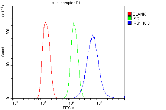
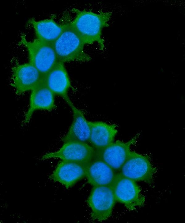
Anti-IRS1 Antibody Picoband™ (monoclonal, 10I3)

- SPECIFICATION
- CITATIONS
- PROTOCOLS
- BACKGROUND

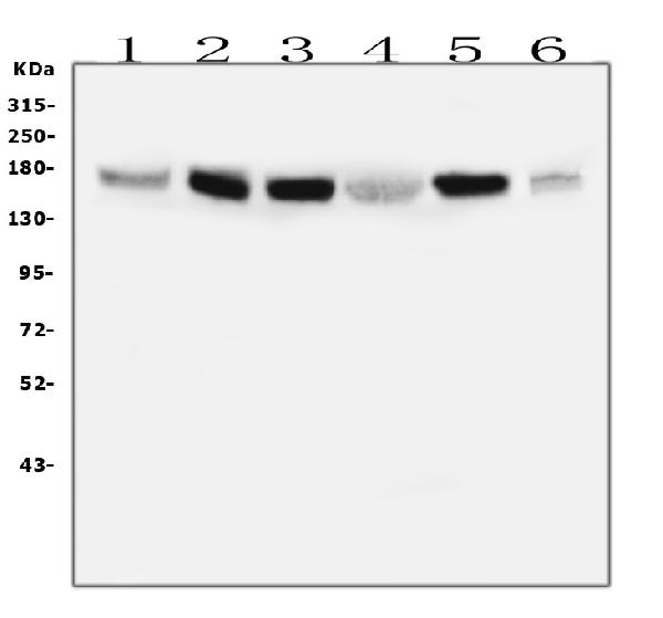
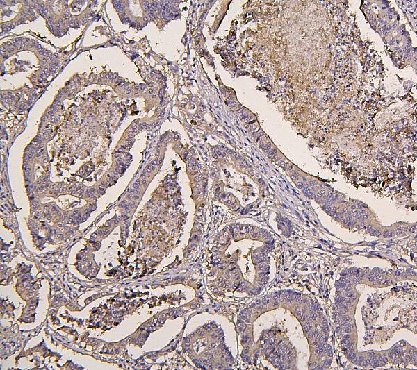
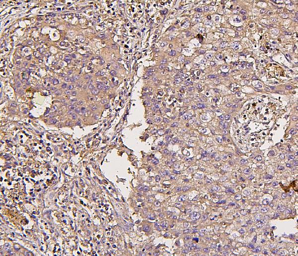
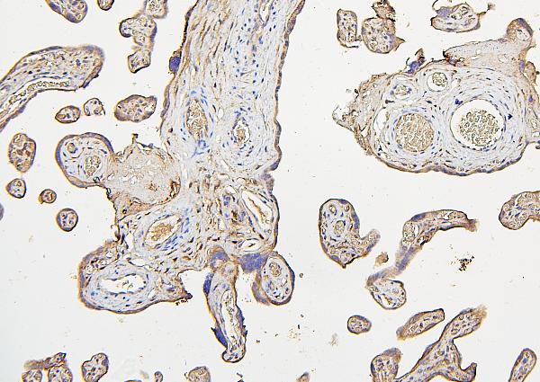
Application
| WB, IHC, IF, ICC, FC |
|---|---|
| Primary Accession | P35568 |
| Host | Mouse |
| Isotype | Mouse IgG2a |
| Reactivity | Human |
| Clonality | Monoclonal |
| Format | Lyophilized |
| Description | Anti-IRS1 Antibody Picoband™ (monoclonal, 10I3) . Tested in Flow Cytometry, IF, IHC, ICC, WB applications. This antibody reacts with Human. |
| Reconstitution | Add 0.2ml of distilled water will yield a concentration of 500 µg/ml. |
| Gene ID | 3667 |
|---|---|
| Other Names | Insulin receptor substrate 1, IRS-1, IRS1 |
| Calculated MW | 160-180 kDa |
| Application Details | Western blot, 0.1-0.5 µg/ml Immunohistochemistry (Paraffin-embedded Section), 0.5-1 µg/ml Immunocytochemistry/Immunofluorescence, 2 µg/ml Flow Cytometry, 1-3 µg/1x10^6 cells |
| Subcellular Localization | Cytosol. Nucleus. Caveola. Insulin receptor complex. Plasma membrane. |
| Contents | Each vial contains 4mg Trehalose, 0.9mg NaCl, 0.2mg Na2HPO4, 0.05mg NaN3. |
| Clone Names | Clone: 10I3 |
| Immunogen | E.coli-derived human IRS1 recombinant protein (Position: S1041-Q1242). Human IRS1 shares 78% and 80% amino acid (aa) sequence identity with mouse and rat IRS1, respectively. |
| Cross Reactivity | No cross-reactivity with other proteins. |
| Storage | Store at -20˚C for one year from date of receipt. After reconstitution, at 4˚C for one month. It can also be aliquotted and stored frozen at -20˚C for six months. Avoid repeated freeze-thaw cycles. |
| Name | IRS1 |
|---|---|
| Function | May mediate the control of various cellular processes by insulin. When phosphorylated by the insulin receptor binds specifically to various cellular proteins containing SH2 domains such as phosphatidylinositol 3-kinase p85 subunit or GRB2. Activates phosphatidylinositol 3-kinase when bound to the regulatory p85 subunit (By similarity). |

Thousands of laboratories across the world have published research that depended on the performance of antibodies from Abcepta to advance their research. Check out links to articles that cite our products in major peer-reviewed journals, organized by research category.
info@abcepta.com, and receive a free "I Love Antibodies" mug.

Provided below are standard protocols that you may find useful for product applications.
Background
Insulin receptor substrate 1 (IRS-1) is a signalling adapter protein that in humans is encoded by the IRS-1 gene. It is mapped to 2q36.3. This gene exhibited no intrinsic enzyme activity, and it can serve as a docking protein involved in binding and activating other signal transduction molecules after being phosphorylated on tyrosine by insulin receptor kinase. IRS1 plays a key role in transmitting signals from the insulin and insulin-like growth factor-1 (IGF-1) receptors to intracellular pathways PI3K/Akt and Erk MAP kinase pathways. IRS1 also has important biological function for both metabolic and mitogenic (growth promoting) pathways. In addition to those, IRS1 is a key regulator of PI3K within malignant cells.
If you have used an Abcepta product and would like to share how it has performed, please click on the "Submit Review" button and provide the requested information. Our staff will examine and post your review and contact you if needed.
If you have any additional inquiries please email technical services at tech@abcepta.com.
Ordering Information
Other Products
Shipping Information
