


 Foundational characteristics of cancer include proliferation, angiogenesis, migration, evasion of apoptosis, and cellular immortality. Find key markers for these cellular processes and antibodies to detect them.
 The SUMOplot™ Analysis Program predicts and scores sumoylation sites in your protein. SUMOylation is a post-translational modification involved in various cellular processes, such as nuclear-cytosolic transport, transcriptional regulation, apoptosis, protein stability, response to stress, and progression through the cell cycle.
 The Autophagy Receptor Motif Plotter predicts and scores autophagy receptor binding sites in your protein. Identifying proteins connected to this pathway is critical to understanding the role of autophagy in physiological as well as pathological processes such as development, differentiation, neurodegenerative diseases, stress, infection, and cancer.
Anti-BAK/BAK1 Antibody Picoband™ (monoclonal, 4C2)
- SPECIFICATION
- CITATIONS
- PROTOCOLS
- BACKGROUND

Application
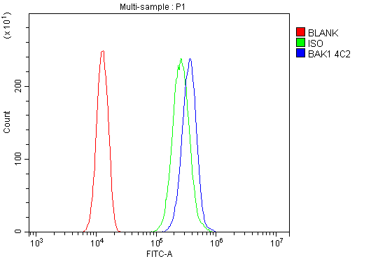
| WB, IHC, IF, ICC, FC |
|---|---|
| Primary Accession | Q16611 |
| Host | Mouse |
| Isotype | Mouse IgG2b |
| Reactivity | Rat, Human, Mouse |
| Clonality | Monoclonal |
| Format | Lyophilized |
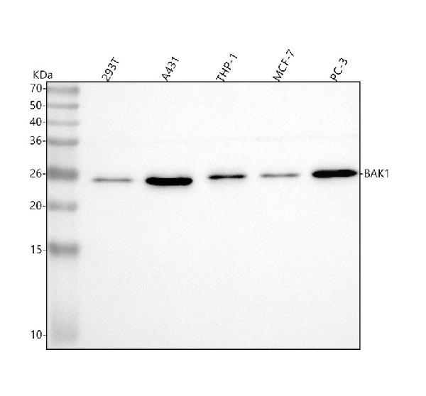
| Description | Anti-BAK/BAK1 Antibody Picoband™ (monoclonal, 4C2) . Tested in Flow Cytometry, IF, IHC, ICC, WB applications. This antibody reacts with Human, Mouse, Rat. |
| Reconstitution | Add 0.2ml of distilled water will yield a concentration of 500 µg/ml. |
| Gene ID | 578 |
|---|---|
| Other Names | Bcl-2 homologous antagonist/killer, Apoptosis regulator BAK, Bcl-2-like protein 7, Bcl2-L-7, BAK1, BAK, BCL2L7, CDN1 |
| Calculated MW | 23-25 kDa |
| Application Details | Western blot, 0.1-0.5 µg/ml Immunohistochemistry (Paraffin-embedded Section), 0.5-1 µg/ml Immunocytochemistry/Immunofluorescence, 2 µg/ml Flow Cytometry, 1-3 µg/1x10^6 cells |
| Subcellular Localization | Mitochondrion outer membrane. Single-pass membrane protein. |
| Tissue Specificity | Expressed in a wide variety of tissues, with highest levels in the heart and skeletal muscle. |
| Contents | Each vial contains 4mg Trehalose, 0.9mg NaCl, 0.2mg Na2HPO4, 0.05mg NaN3. |
| Clone Names | Clone: 4C2 |
| Immunogen | E.coli-derived human BAK/BAK1 recombinant protein (Position: A22-S211). Human BAK shares 78.3 % amino acid (aa) sequence identity with mouse BAK. |
| Cross Reactivity | No cross-reactivity with other proteins. |
| Storage | Store at -20˚C for one year from date of receipt. After reconstitution, at 4˚C for one month. It can also be aliquotted and stored frozen at -20˚C for six months. Avoid repeated freeze-thaw cycles. |
| Name | BAK1 |
|---|---|
| Synonyms | BAK, BCL2L7, CDN1 |
| Function | Plays a role in the mitochondrial apoptotic process. Upon arrival of cell death signals, promotes mitochondrial outer membrane (MOM) permeabilization by oligomerizing to form pores within the MOM. This releases apoptogenic factors into the cytosol, including cytochrome c, promoting the activation of caspase 9 which in turn processes and activates the effector caspases. |
| Cellular Location | Mitochondrion outer membrane; Single-pass membrane protein |
| Tissue Location | Expressed in a wide variety of tissues, with highest levels in the heart and skeletal muscle |

Thousands of laboratories across the world have published research that depended on the performance of antibodies from Abcepta to advance their research. Check out links to articles that cite our products in major peer-reviewed journals, organized by research category.
info@abcepta.com, and receive a free "I Love Antibodies" mug.

Provided below are standard protocols that you may find useful for product applications.
Background
BAK, officially called Bcl2 antagonist killer, is a protein that in humans, encoded by the BAK gene. The BAK protein is a pro-apoptotic member of the Bcl-2 gene family which is involved in initiating apoptosis. BAK gene spans 7.6 kb and contains 6 exons. By Southern blot analysis of genomic DNA from human/rodent somatic cell hybrids, BAK gene is localized to chromosome 6. This protein localizes to mitochondria, and functions to induce apoptosis. It interacts with and accelerates the opening of the mitochondrial voltage-dependent anion channel, which leads to a loss in membrane potential and the release of cytochrome. This protein also interacts with the tumor suppressor P53 after exposure to cell stress.
If you have used an Abcepta product and would like to share how it has performed, please click on the "Submit Review" button and provide the requested information. Our staff will examine and post your review and contact you if needed.
If you have any additional inquiries please email technical services at tech@abcepta.com.
Ordering Information
Other Products
Shipping Information
