


 Foundational characteristics of cancer include proliferation, angiogenesis, migration, evasion of apoptosis, and cellular immortality. Find key markers for these cellular processes and antibodies to detect them.
 The SUMOplot™ Analysis Program predicts and scores sumoylation sites in your protein. SUMOylation is a post-translational modification involved in various cellular processes, such as nuclear-cytosolic transport, transcriptional regulation, apoptosis, protein stability, response to stress, and progression through the cell cycle.
 The Autophagy Receptor Motif Plotter predicts and scores autophagy receptor binding sites in your protein. Identifying proteins connected to this pathway is critical to understanding the role of autophagy in physiological as well as pathological processes such as development, differentiation, neurodegenerative diseases, stress, infection, and cancer.
Anti-Cofilin 2/CFL2 Antibody Picoband™ (monoclonal, 8C13)
- SPECIFICATION
- CITATIONS
- PROTOCOLS
- BACKGROUND

Application
| WB, IHC, IF, ICC, FC |
|---|---|
| Primary Accession | Q9Y281 |
| Host | Mouse |
| Isotype | Mouse IgG2b |
| Reactivity | Rat, Human, Mouse |
| Clonality | Monoclonal |
| Format | Lyophilized |
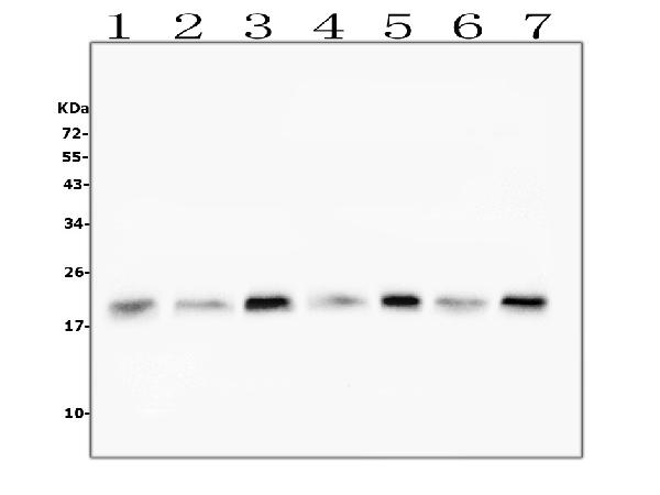
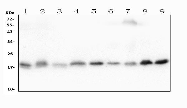
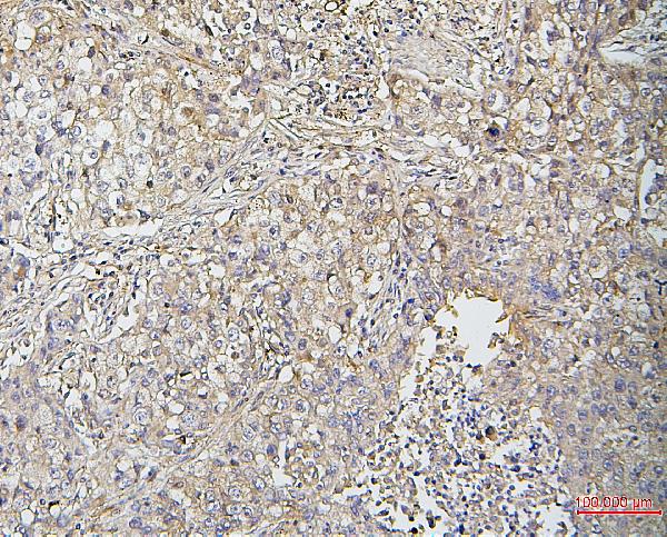
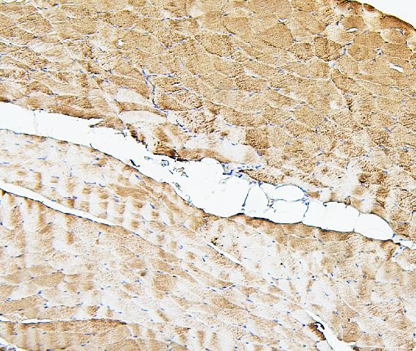
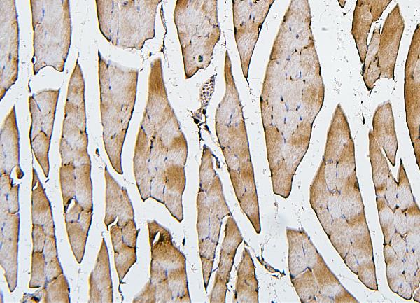
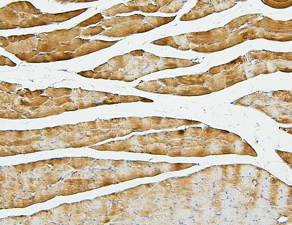
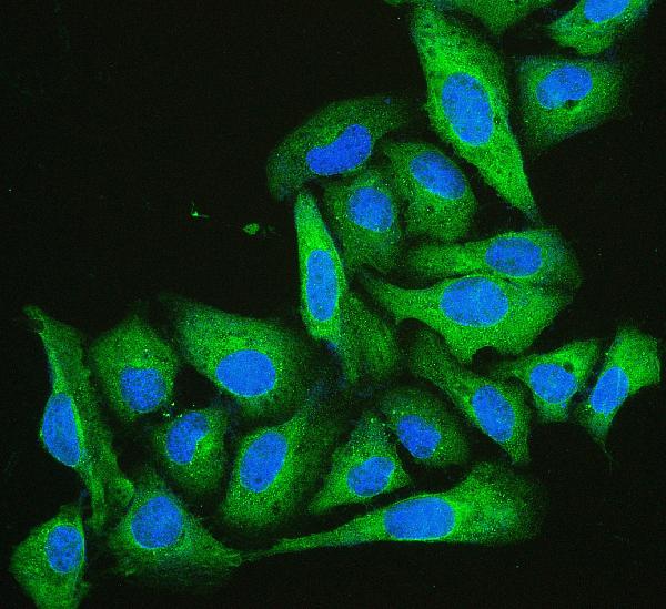
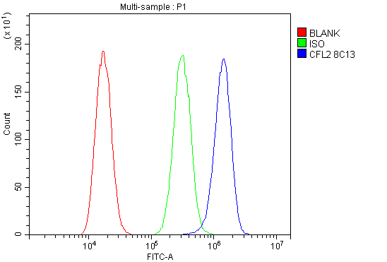
| Description | Anti-Cofilin 2/CFL2 Antibody Picoband™ (monoclonal, 8C13) . Tested in Flow Cytometry, IF, IHC, ICC, WB applications. This antibody reacts with Human, Mouse, Rat. |
| Reconstitution | Add 0.2ml of distilled water will yield a concentration of 500 µg/ml. |
| Gene ID | 1073 |
|---|---|
| Other Names | Cofilin-2, Cofilin, muscle isoform, CFL2 |
| Calculated MW | 19 kDa |
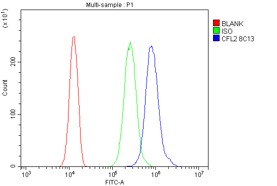
| Application Details | Western blot, 0.1-0.5 µg/ml Immunohistochemistry (Paraffin-embedded Section), 0.5-1 µg/ml Immunocytochemistry/Immunofluorescence, 2 µg/ml Flow Cytometry, 1-3 µg/1x10^6 cells |
| Subcellular Localization | Cytoskeleton. Nucleus matrix. |
| Tissue Specificity | Isoform CFL2b is expressed predominantly in skeletal muscle and heart. Isoform CFL2a is expressed in various tissues. |
| Contents | Each vial contains 4mg Trehalose, 0.9mg NaCl, 0.2mg Na2HPO4, 0.05mg NaN3. |
| Clone Names | Clone: 8C13 |
| Immunogen | A synthetic peptide corresponding to a sequence at the C-terminus of human Cofilin 2/CFL2, identical to the related mouse sequence. |
| Cross Reactivity | No cross-reactivity with other proteins. |
| Storage | Store at -20˚C for one year from date of receipt. After reconstitution, at 4˚C for one month. It can also be aliquotted and stored frozen at -20˚C for six months. Avoid repeated freeze-thaw cycles. |
| Name | CFL2 |
|---|---|
| Function | Controls reversibly actin polymerization and depolymerization in a pH-sensitive manner. Its F-actin depolymerization activity is regulated by association with CSPR3 (PubMed:19752190). It has the ability to bind G- and F-actin in a 1:1 ratio of cofilin to actin. It is the major component of intranuclear and cytoplasmic actin rods. Required for muscle maintenance. May play a role during the exchange of alpha-actin forms during the early postnatal remodeling of the sarcomere (By similarity). |
| Cellular Location | Nucleus matrix. Cytoplasm, cytoskeleton. Note=Colocalizes with CSPR3 in the Z line of sarcomeres. |
| Tissue Location | Isoform CFL2b is expressed predominantly in skeletal muscle and heart. Isoform CFL2a is expressed in various tissues |

Thousands of laboratories across the world have published research that depended on the performance of antibodies from Abcepta to advance their research. Check out links to articles that cite our products in major peer-reviewed journals, organized by research category.
info@abcepta.com, and receive a free "I Love Antibodies" mug.

Provided below are standard protocols that you may find useful for product applications.
Background
Cofilin 2 (muscle), also known as CFL2, is a protein which in humans is encoded by the CFL2 gene. It is mapped to 14q12. This gene encodes an intracellular protein that is involved in the regulation of actin-filament dynamics. And this protein is a major component of intranuclear and cytoplasmic actin rods. It can bind G- and F-actin in a 1:1 ratio of cofilin to actin, and it reversibly controls actin polymerization and depolymerization in a pH-dependent manner. Mutations in this gene cause nemaline myopathy type 7, a form of congenital myopathy. Alternative splicing results in multiple transcript variants.
If you have used an Abcepta product and would like to share how it has performed, please click on the "Submit Review" button and provide the requested information. Our staff will examine and post your review and contact you if needed.
If you have any additional inquiries please email technical services at tech@abcepta.com.
Ordering Information
Other Products
Shipping Information
