


 Foundational characteristics of cancer include proliferation, angiogenesis, migration, evasion of apoptosis, and cellular immortality. Find key markers for these cellular processes and antibodies to detect them.
 The SUMOplot™ Analysis Program predicts and scores sumoylation sites in your protein. SUMOylation is a post-translational modification involved in various cellular processes, such as nuclear-cytosolic transport, transcriptional regulation, apoptosis, protein stability, response to stress, and progression through the cell cycle.
 The Autophagy Receptor Motif Plotter predicts and scores autophagy receptor binding sites in your protein. Identifying proteins connected to this pathway is critical to understanding the role of autophagy in physiological as well as pathological processes such as development, differentiation, neurodegenerative diseases, stress, infection, and cancer.
Anti-NMI Antibody Picoband™ (monoclonal, 2F3)
- SPECIFICATION
- CITATIONS
- PROTOCOLS
- BACKGROUND

Application
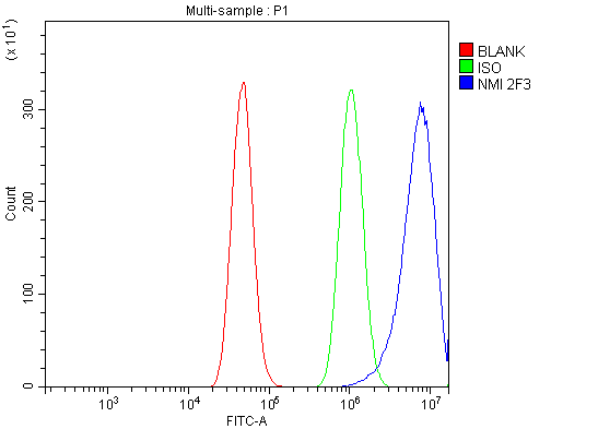
| WB, IHC, IF, ICC, FC |
|---|---|
| Primary Accession | Q13287 |
| Host | Mouse |
| Isotype | Mouse IgG2b |
| Reactivity | Rat, Human, Mouse |
| Clonality | Monoclonal |
| Format | Lyophilized |
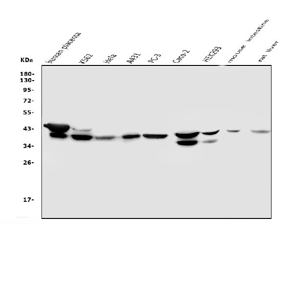
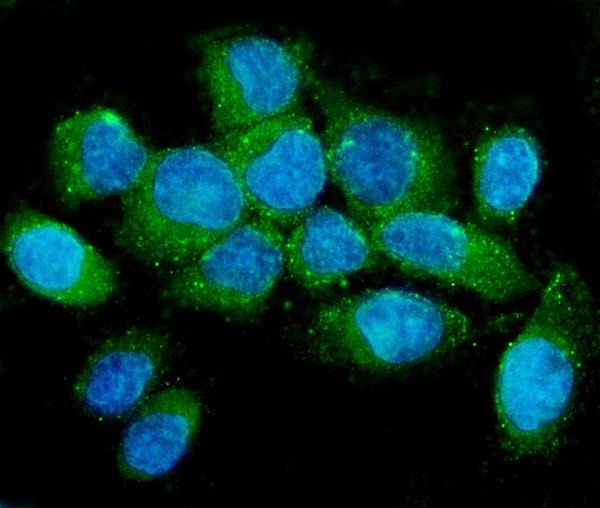
| Description | Anti-NMI Antibody Picoband™ (monoclonal, 2F3) . Tested in Flow Cytometry, IF, IHC, ICC, WB applications. This antibody reacts with Human, Mouse, Rat. |
| Reconstitution | Add 0.2ml of distilled water will yield a concentration of 500 µg/ml. |
| Gene ID | 9111 |
|---|---|
| Other Names | N-myc-interactor, Nmi, N-myc and STAT interactor, NMI (HGNC:7854) |
| Calculated MW | 40 kDa |
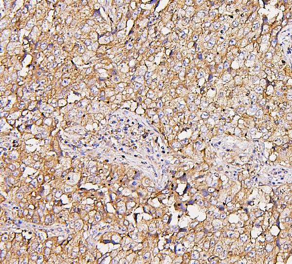
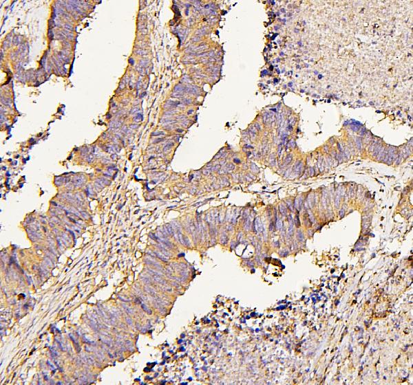
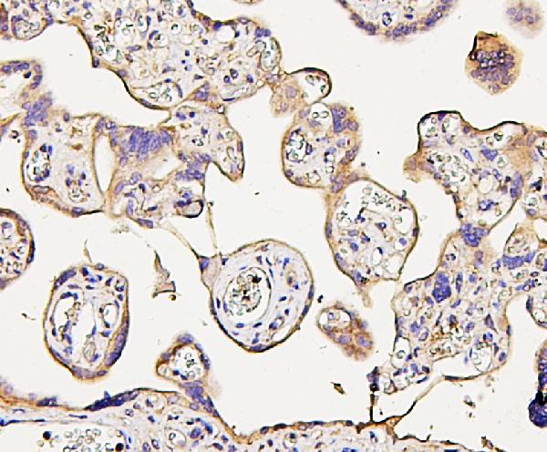
| Application Details | Western blot, 0.1-0.5 µg/ml Immunohistochemistry (Paraffin-embedded Section), 0.5-1 µg/ml Immunocytochemistry/Immunofluorescence, 5 µg/ml Flow Cytometry, 1-3 µg/1x10^6 cells |
| Subcellular Localization | Cytoplasm. |
| Tissue Specificity | Expressed in all adult and fetal tissues except brain and skin. More abundant in fetal tissues especially liver. |
| Contents | Each vial contains 4mg Trehalose, 0.9mg NaCl, 0.2mg Na2HPO4, 0.05mg NaN3. |
| Clone Names | Clone: 2F3 |
| Immunogen | E.coli-derived human NMI recombinant protein (Position: E2-E307). Human NMI shares 64% amino acid (aa) sequence identity with mouse NMI. |
| Cross Reactivity | No cross-reactivity with other proteins. |
| Storage | Store at -20˚C for one year from date of receipt. After reconstitution, at 4˚C for one month. It can also be aliquotted and stored frozen at -20˚C for six months. Avoid repeated freeze-thaw cycles. |
| Name | NMI (HGNC:7854) |
|---|---|
| Function | Acts as a signaling pathway regulator involved in innate immune system response (PubMed:26342464, PubMed:29038465, PubMed:29350881, PubMed:9989503). In response to interleukin 2/IL2 and interferon IFN-gamma/IFNG, interacts with signal transducer and activator of transcription/STAT which activate the transcription of downstream genes involved in a multitude of signals for development and homeostasis (PubMed:29377960, PubMed:9989503). Enhances the recruitment of CBP/p300 coactivators to STAT1 and STAT5, resulting in increased STAT1- and STAT5-dependent transcription (PubMed:9989503). In response to interferon IFN-alpha, associates in a complex with signaling pathway regulator IFI35 to regulate immune response; the complex formation prevents proteasome-mediated degradation of IFI35 (PubMed:10779520, PubMed:10950963). In complex with IFI35, inhibits virus-triggered type I IFN-beta production when ubiquitinated by ubiquitin-protein ligase TRIM21 (PubMed:26342464). In complex with IFI35, negatively regulates nuclear factor NF-kappa-B signaling by inhibiting the nuclear translocation, activation and transcription of NF-kappa-B subunit p65/RELA, resulting in the inhibition of endothelial cell proliferation, migration and re-endothelialization of injured arteries (PubMed:29350881). Negatively regulates virus-triggered type I interferon/IFN production by inducing proteosome-dependent degradation of IRF7, a transcriptional regulator of type I IFN, thereby interfering with cellular antiviral responses (By similarity). Beside its role as an intracellular signaling pathway regulator, also functions extracellularly as damage-associated molecular patterns (DAMPs) to promote inflammation, when actively released by macrophage to the extracellular space during cell injury or pathogen invasion (PubMed:29038465). Macrophage-secreted NMI activates NF-kappa-B signaling in adjacent macrophages through Toll-like receptor 4/TLR4 binding and activation, thereby inducing NF-kappa-B translocation from the cytoplasm into the nucleus which promotes the release of pro- inflammatory cytokines (PubMed:29038465). |
| Cellular Location | Cytoplasm. Nucleus. Secreted Note=Cytoplasmic NMI localizes in punctate granular structures (PubMed:10950963, PubMed:9781816). Nuclear localization increased following IFN-alpha treatment (PubMed:10950963, PubMed:9781816) Extracelullar following secretion by macrophage (PubMed:29038465) |
| Tissue Location | Expressed in adult spleen, liver, and kidney (PubMed:9781816). Expressed in fetal thymus, liver, placenta, spleen, lung, and kidney but not brain (PubMed:9781816). Expressed in macrophages (PubMed:29038465). |

Thousands of laboratories across the world have published research that depended on the performance of antibodies from Abcepta to advance their research. Check out links to articles that cite our products in major peer-reviewed journals, organized by research category.
info@abcepta.com, and receive a free "I Love Antibodies" mug.

Provided below are standard protocols that you may find useful for product applications.
Background
NMYC interactor (NMI) encodes a protein that interacts with NMYC and CMYC (two members of the oncogene Myc family), and other transcription factors containing a Zip, HLH, or HLH-Zip motif. The NMI protein also interacts with all STATs except STAT2 and augments STAT-mediated transcription in response to cytokines IL2 and IFN-gamma. The NMI mRNA has low expression levels in all human fetal and adult tissues tested except brain and has high expression in cancer cell line-myeloid leukemias.
If you have used an Abcepta product and would like to share how it has performed, please click on the "Submit Review" button and provide the requested information. Our staff will examine and post your review and contact you if needed.
If you have any additional inquiries please email technical services at tech@abcepta.com.
Ordering Information
Other Products
Shipping Information
