


 Foundational characteristics of cancer include proliferation, angiogenesis, migration, evasion of apoptosis, and cellular immortality. Find key markers for these cellular processes and antibodies to detect them.
 The SUMOplot™ Analysis Program predicts and scores sumoylation sites in your protein. SUMOylation is a post-translational modification involved in various cellular processes, such as nuclear-cytosolic transport, transcriptional regulation, apoptosis, protein stability, response to stress, and progression through the cell cycle.
 The Autophagy Receptor Motif Plotter predicts and scores autophagy receptor binding sites in your protein. Identifying proteins connected to this pathway is critical to understanding the role of autophagy in physiological as well as pathological processes such as development, differentiation, neurodegenerative diseases, stress, infection, and cancer.
Anti-Collagen III/COL3A1 Antibody Picoband™ (monoclonal, 9H9)
- SPECIFICATION
- CITATIONS
- PROTOCOLS
- BACKGROUND

Application
| WB, IHC |
|---|---|
| Primary Accession | P02461 |
| Host | Mouse |
| Isotype | Mouse IgG2b |
| Reactivity | Rat, Human, Mouse |
| Clonality | Monoclonal |
| Format | Lyophilized |
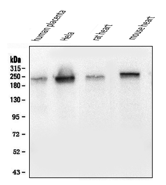
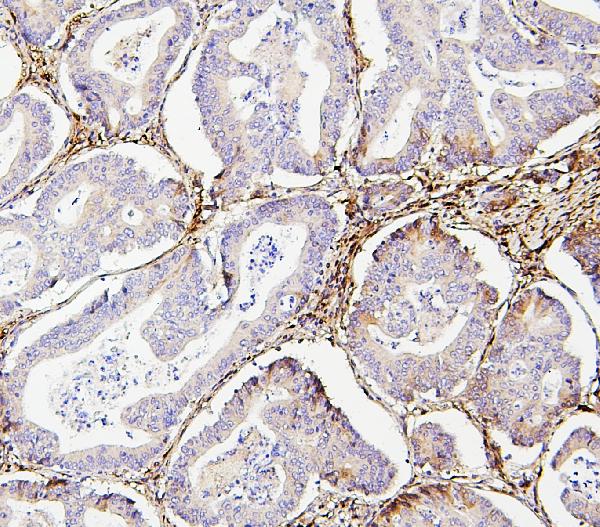
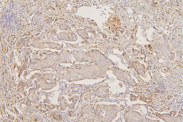
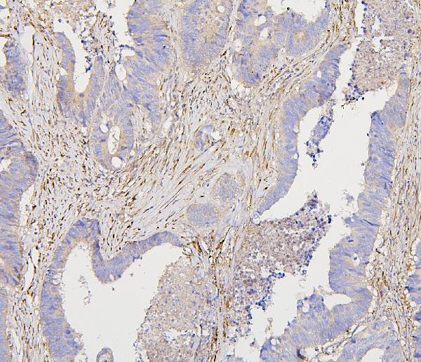
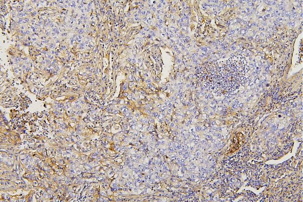
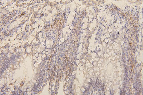
| Description | Anti-Collagen III/COL3A1 Antibody Picoband™ (monoclonal, 9H9) . Tested in IHC, WB applications. This antibody reacts with Human, Mouse, Rat. |
| Reconstitution | Add 0.2ml of distilled water will yield a concentration of 500 µg/ml. |
| Gene ID | 1281 |
|---|---|
| Other Names | Collagen alpha-1(III) chain, COL3A1 |
| Calculated MW | 180-190 kDa |
| Application Details | Western blot, 0.1-0.5 µg/ml Immunohistochemistry (Paraffin-embedded Section), 0.5-1 µg/ml |
| Subcellular Localization | Extracellular matrix. |
| Contents | Each vial contains 4mg Trehalose, 0.9mg NaCl, 0.2mg Na2HPO4, 0.05mg NaN3. |
| Clone Names | Clone: 9H9 |
| Immunogen | E. coli-derived human Collagen III/COL3A1 recombinant protein (Position: D1222- E1455). |
| Cross Reactivity | No cross-reactivity with other proteins. |
| Storage | Store at -20˚C for one year from date of receipt. After reconstitution, at 4˚C for one month. It can also be aliquotted and stored frozen at -20˚C for six months. Avoid repeated freeze-thaw cycles. |
| Name | COL3A1 |
|---|---|
| Function | Collagen type III occurs in most soft connective tissues along with type I collagen. Involved in regulation of cortical development. Is the major ligand of ADGRG1 in the developing brain and binding to ADGRG1 inhibits neuronal migration and activates the RhoA pathway by coupling ADGRG1 to GNA13 and possibly GNA12. |
| Cellular Location | Secreted, extracellular space, extracellular matrix {ECO:0000255|PROSITE-ProRule:PRU00793} |

Thousands of laboratories across the world have published research that depended on the performance of antibodies from Abcepta to advance their research. Check out links to articles that cite our products in major peer-reviewed journals, organized by research category.
info@abcepta.com, and receive a free "I Love Antibodies" mug.

Provided below are standard protocols that you may find useful for product applications.
Background
COL3A1, also called EDS4A or Collagen alpha-1 (III), is a protein that in humans is encoded by the COL3A1 gene. It is mapped to 2q32.2. COL3A1 chain is a fibrillar-forming collagen comprising 3 alpha-1 (III) chains and is expressed in early embryos and throughout embryogenesis. In adult, COL3A1 is a major component of the extracellular matrix in a variety of internal organs and skin. COL3A1 is also a fibrous scleroprotein in bone, cartilage, dentin, tendon, bone marrow stroma and other connective tissue. It is involved in regulation of cortical development, and it is the major ligand of GPR56 in the developing brain. COL3A1 binding to GPR56 can inhibit neuronal migration and activate the RhoA pathway by coupling GPR56 to GNA13 and possibly GNA12.
If you have used an Abcepta product and would like to share how it has performed, please click on the "Submit Review" button and provide the requested information. Our staff will examine and post your review and contact you if needed.
If you have any additional inquiries please email technical services at tech@abcepta.com.
Ordering Information
Other Products
Shipping Information
