


 Foundational characteristics of cancer include proliferation, angiogenesis, migration, evasion of apoptosis, and cellular immortality. Find key markers for these cellular processes and antibodies to detect them.
 The SUMOplot™ Analysis Program predicts and scores sumoylation sites in your protein. SUMOylation is a post-translational modification involved in various cellular processes, such as nuclear-cytosolic transport, transcriptional regulation, apoptosis, protein stability, response to stress, and progression through the cell cycle.
 The Autophagy Receptor Motif Plotter predicts and scores autophagy receptor binding sites in your protein. Identifying proteins connected to this pathway is critical to understanding the role of autophagy in physiological as well as pathological processes such as development, differentiation, neurodegenerative diseases, stress, infection, and cancer.
Anti-ATP citrate lyase ACLY Antibody Picoband™ (monoclonal, 5I2)
- SPECIFICATION
- CITATIONS
- PROTOCOLS
- BACKGROUND

Application
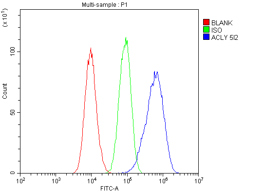
| WB, IHC, IF, ICC, FC |
|---|---|
| Primary Accession | P53396 |
| Host | Mouse |
| Isotype | Mouse IgG2b |
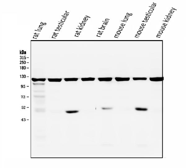
| Reactivity | Rat, Human, Mouse |
| Clonality | Monoclonal |
| Format | Lyophilized |
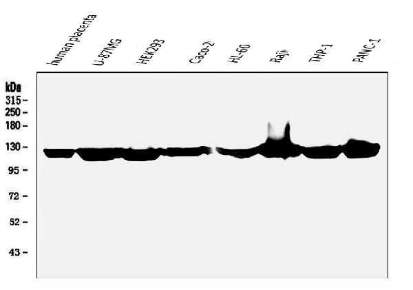
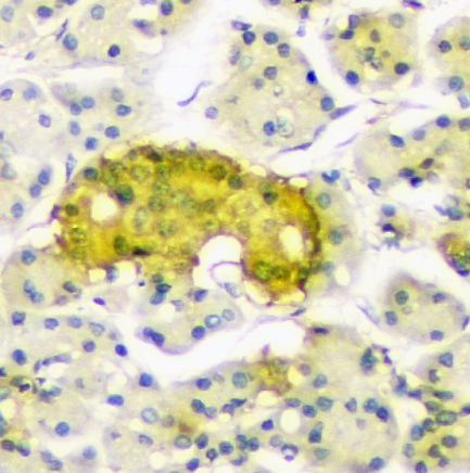
| Description | Anti-ATP citrate lyase ACLY Antibody Picoband™ (monoclonal, 5I2) . Tested in Flow Cytometry, IF, IHC, ICC, WB applications. This antibody reacts with Human, Mouse, Rat. |
| Reconstitution | Add 0.2ml of distilled water will yield a concentration of 500 µg/ml. |
| Gene ID | 47 |
|---|---|
| Other Names | ATP-citrate synthase, 2.3.3.8, ATP-citrate (pro-S-)-lyase, ACL, Citrate cleavage enzyme, ACLY |
| Calculated MW | 121 kDa |
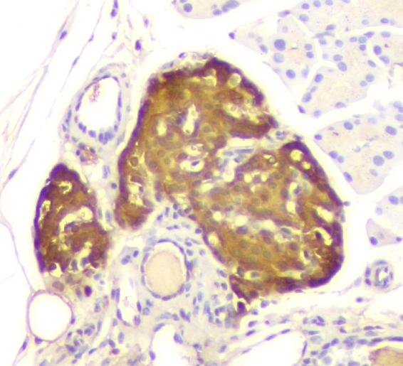
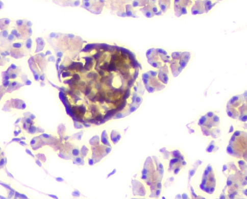
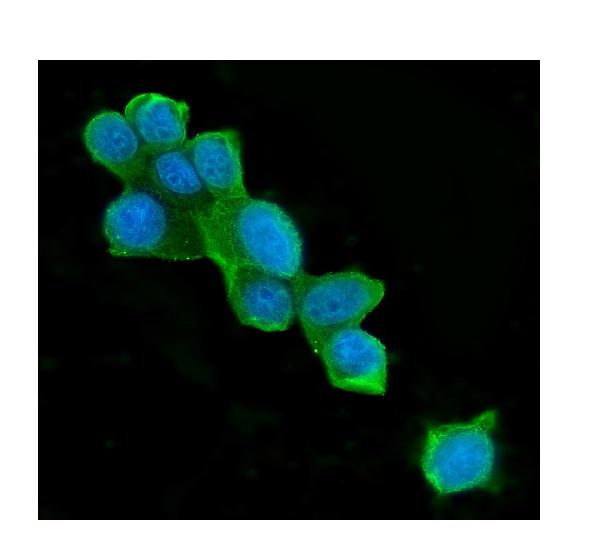
| Application Details | Western blot, 0.1-0.5 µg/ml Immunohistochemistry (Paraffin-embedded Section), 0.5-1 µg/ml Immunocytochemistry/Immunofluorescence, 2 µg/ml, Human Flow Cytometry, 1-3 µg/1x10^6 cells |
| Subcellular Localization | Cytoplasm. |
| Contents | Each vial contains 4mg Trehalose, 0.9mg NaCl, 0.2mg Na2HPO4, 0.05mg NaN3. |
| Clone Names | Clone: 5I2 |
| Immunogen | E. coli-derived human ATP citrate lyase recombinant protein (Position: M1-I180). Human ATP citrate lyase shares 95% amino acid (aa) sequence identity with both mouse and rat ATP citrate lyase. |
| Cross Reactivity | No cross-reactivity with other proteins. |
| Storage | Store at -20˚C for one year from date of receipt. After reconstitution, at 4˚C for one month. It can also be aliquotted and stored frozen at -20˚C for six months. Avoid repeated freeze-thaw cycles. |
| Name | ACLY |
|---|---|
| Function | Catalyzes the cleavage of citrate into oxaloacetate and acetyl-CoA, the latter serving as common substrate for de novo cholesterol and fatty acid synthesis. |
| Cellular Location | Cytoplasm, cytosol. |

Thousands of laboratories across the world have published research that depended on the performance of antibodies from Abcepta to advance their research. Check out links to articles that cite our products in major peer-reviewed journals, organized by research category.
info@abcepta.com, and receive a free "I Love Antibodies" mug.

Provided below are standard protocols that you may find useful for product applications.
Background
ATP citrate lyase, aslo known as ACLY, is an enzyme that in animals represents an important step in fatty acid biosynthesis. ATP citrate lyase is the primary enzyme responsible for the synthesis of cytosolic acetyl-CoA in many tissues. The enzyme is a tetramer of apparently identical subunits. The product, acetyl-CoA, in animals serves several important biosynthetic pathways, including lipogenesis and cholesterogenesis. It is activated by insulin. In nervous tissue, ATP citrate-lyase may be involved in the biosynthesis of acetylcholine.In plants, ATP citrate lyase generates the acetyl-CoA for cytosolically-synthesized metabolites.
If you have used an Abcepta product and would like to share how it has performed, please click on the "Submit Review" button and provide the requested information. Our staff will examine and post your review and contact you if needed.
If you have any additional inquiries please email technical services at tech@abcepta.com.
Ordering Information
Other Products
Shipping Information
